Can lead placement affect ECG
Deviation of lead placement even by 20-25mm from the correct position can create clinically significant changes on the ECG, including changes to the ST-segment (McCann et al. 2007).
Can incorrect lead placement affect ECG?
The analysis of ECG signals recorded from misplaced electrodes can lead to misinterpretation or even to significant diagnostic errors like incorrect recognition of anterior infarction, anteroseptal infarction, ventricular hypertrophy [9, 14], false diagnosis of ischemia, or Brugada syndrome [16, 24].
What issues could compromise the quality of ECG tracing?
- Obesity.
- Anatomical considerations, such as the size of the chest and the location of the heart within the chest.
- Movement during the test.
- Exercise or smoking before the test.
- Certain medicines.
- Electrolyte imbalances, such as too much or too little potassium, magnesium, or calcium in the blood.
Why is it important to position electrodes correctly?
The system of positioning of leads for performing a 12-lead ECG is universal. This helps to ensure that, when a person’s ECGs are compared, any changes on the ECG are due to cardiac injury, not a difference in placement of leads, this is extremely important with the increasing use of foreign travel.What happens to an ECG when we use a modified electrode limb placement?
Modified Limb Lead (MLL) ECG system may be used during rest or exercise ECG, or atrial activity enhancement. Because of modification in the limb electrode placement, changes are likely to happen in ECG wave amplitudes and frontal plane axis, which may alter the clinical limits of normality and ECG diagnostic criteria.
Can ECG be wrong?
The study of 500 patients found a false positive reading between 77 and 82 percent in patients screened by electrocardiogram, and a false negative reading between 6 percent to 7 percent in the same patient population.
How do I know if my ECG is wrong?
The mistake can be recognized by the presence of unusual P–QRS patterns (e.g. negative P–QRS in lead I or II, positive in lead AVR, P–QRS complexes of opposite direction in leads I and V6, etc.), change in the P–QRS axis, or abnormal precordial QRS–T wave progression.
Where do female ECG leads go?
For females, place the leads under the breast tissue. You may need to lift and clean the skin underneath the breast to get a clear tracing. A quality 12-lead ECG has a smooth, flat baseline (called the isoelectric line).Where do heart monitor leads go?
RA placementWHITE directly below the clavicle and near the right shoulder.LL placementRED on the left lower abdomen.V placementBROWN on the chest, the position depends on your required lead selection (4th intercostal space, the right side of the sternum).
Where do ECG leads go on a dog?Electrodes should be placed: Red = right forelimb, placed behind the elbow. Yellow = left forelimb, placed behind the elbow. Green = left hindlimb, placed at the front of the stifle.
Article first time published onWhat can cause interference with an ECG?
Electromagnetic interference (EMI) artifact usually results from electrical power lines, electrical equipment, and mobile telephones. In the United States this is sometimes referred to as 60 cycle interference (or 60 Hz pickup).
What could interfere with performing electrocardiography?
Artefact Interference that may impact accurate interpretation. Common examples include: 60 Hz cycle interference, muscle tremor, wandering baseline, poor electrode contact, patient movement, improper standardisation and limb lead reversal.
Can anxiety affect ECG results?
Premature ventricular contractions is one of the manifestations of sympathetic over activity due to anxiety. However, anxiety might induce electrocardiographic (ECG) changes in normal person with normal heart, as in this documented case.
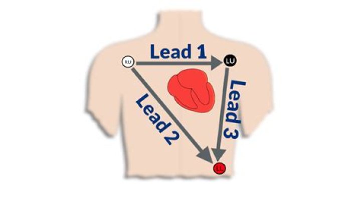
What are limb Leads in ECG?
Limb leads are made up of 4 leads placed on the extremities: left and right wrist; left and right ankle. The lead connected to the right ankle is a neutral lead, like you would find in an electric plug. It is there to complete an electrical circuit and plays no role in the ECG itself.
Where do you place a 3 lead ECG?
- WHITE.
- RA (right arm), just below the right clavicle.
- BLACK.
- LA (left arm), just below the left clavicle.
- RED.
- LL (left leg), on the lower chest, just above and left of the umbilicus.
Does limb lead placement matter?
Conclusions: We provide better and more robust evidence that routine modification of limb electrode placement produces only minor changes to the ECG waveform in healthy subjects. These are not clinically significant according to the 2009 guidelines and thus have no effect on the clinical specificity of the 12 lead ECG.
Does Gas affect ECG?
Visceral-cardiac reflex secondary to gastric distention which causes increased vagal tone can lead to ECG changes.
Can ECG detect heart blockage?
However, it does not show whether you have asymptomatic blockages in your heart arteries or predict your risk of a future heart attack. The resting ECG is different from a stress or exercise ECG or cardiac imaging test.
Why is ECG lead placement important?
It is important an ECG is recorded accurately. ECG electrode placement is standardised, allowing for the recording of an accurate trace – but also ensuring comparability between records taken at different times.
What causes chest pain if ECG is normal?
It could be a lung disorder, such as a blood clot to the lungs, known as a pulmonary embolism. Additionally, other causes of chest discomfort include spasm of the esophagus, diseases of the aorta, gastroesophageal reflux disease, musculoskeletal pain, fast heart rhythm abnormalities and costochondritis.
Where are the 12 leads placed on a patient for an ECG?
Electrode placement for a 12-lead ECG is standard, with leads placed on the left and right arm and left and right leg. Another pair of electrodes is placed between the fourth and fifth ribs on the left and right side of the sternum.
What are the three types of ECG leads?
- Limb Leads (Bipolar)
- Augmented Limb Leads (Unipolar)
- Chest Leads (Unipolar)
How do you remember ECG lead placement?
- Attach the right arm (RA) electrode.
- Attach the left arm (LA) electrode.
- Attach the left leg (LL) electrode.
- Attach the right leg (RL) electrode.
Why it is called 12 lead ECG?
In other words, each ECG lead is computed by analysing the electrical currents detected by several electrodes. The standard ECG – which is referred to as a 12-lead ECG since it includes 12 leads – is obtained using 10 electrodes.
Do you have to remove bra for EKG?
Women may often wear a bra, T-shirt, or gown. If you are wearing stockings, you should take them off. You will be given a cloth or paper covering to use during the test. Talk to your doctor about any concerns you have regarding the need for the test, its risks, how it will be done, or what the results will mean.
How is ECG done in female?
Generally, the test involves attaching a number of small, sticky sensors called electrodes to your arms, legs and chest. These are connected by wires to an ECG recording machine. You don’t need to do anything special to prepare for the test. You can eat and drink as normal beforehand.
Why does my dog need an ECG?
Why might my pet need one? An ECG is used to identify and diagnose many problems with your pet’s heart. Your vet might suggest your pet has as ECG if they suspect a heart murmur, if an x-ray shows evidence of heart enlargement, or if your pet is showing cardiovascular symptoms such as fainting.
Why would a dog need an ECG?
The electrocardiogram will give your veterinarian a snapshot of electrical rhythm in your dog’s heart. This test will reveal any abnormalities or arrhythmias in the heart. Electrical problems within your dog’s heart can be a symptom of diseases and can also cause irregular heartbeats, fatigue, and lethargy.
In what position is an animal placed for an ECG?
Recording an ECG The ECG detects the electrical activity of the heart through 3 electrodes. In small animals these electrodes are most commonly placed on the 2 forelimbs and the left hindlimb. An additional electrode may be placed on the right hindlimb.
How do you remove motion artifacts from ECG?
An existing method to remove the motion artefact is to employ an accelerometer for measuring the body movement at the same time of ECG detection [5]. However, for non-contact electrode structure of ECG detection, an accelerometer directly attached to the human body is unacceptable.
How often should ECG leads be changed?
Electrodes should be changed daily. Electrode placement is integral for accurate results. When an electrode is misplaced by as little as one intercostal space, QRS morphology may change and contribute to misdiagnosis.