What are sutures and fontanelles
Joints made of strong, fibrous tissue (cranial sutures) hold the bones of your baby’s skull together. The sutures meet at the fontanels, the soft spots on your baby’s head. The sutures remain flexible during infancy, allowing the skull to expand as the brain grows.
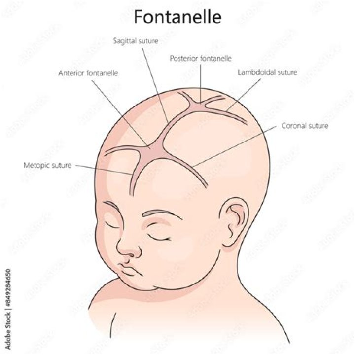
What is a fontanelle?
In an infant, the space where 2 sutures join forms a membrane-covered “soft spot” called a fontanelle (fontanel). The fontanelles allow for growth of the brain and skull during an infant’s first year. There are normally several fontanelles on a newborn’s skull.
What are sutures in a newborn?
Sutures allow the bones to move during the birth process. They act like an expansion joint. This allows the bone to enlarge evenly as the brain grows and the skull expands. The result is a symmetrically shaped head.
What are the sutures?
In anatomy, a suture is a fairly rigid joint between two or more hard elements of an organism, with or without significant overlap of the elements. Sutures are found in the skeletons or exoskeletons of a wide range of animals, in both invertebrates and vertebrates.What are sutures in fetal skull?
Sutures are joints between the bones of the skull. In the fetus they can ‘give’ a little under the pressure on the baby’s head as it passes down the birth canal. … The lambdoid suture forms the junction between the occipital and the frontal bone.
What are the 4 fontanelles?
- Anterior Fontanelle. The anterior fontanelle is the largest of the six fontanelles, and it resembles a diamond-shape ranging in size from 0.6 cm to 3.6 cm with a mean of 2.1 cm. …
- Posterior Fontanelle. …
- Mastoid Fontanelle. …
- Sphenoid Fontanelle. …
- Third Fontanel.
Do fontanelles become sutures?
Joints made of strong, fibrous tissue (cranial sutures) hold the bones of your baby’s skull together. The sutures meet at the fontanels, the soft spots on your baby’s head. The sutures remain flexible during infancy, allowing the skull to expand as the brain grows.
Where is a suture?
A suture is a type of fibrous joint (or synarthrosis) that only occurs in the skull. The bones are bound together by Sharpey’s fibers, a matrix of connective tissue which provide a firm joint.What are the 3 types of sutures?
- Continuous sutures. This technique involves a series of stitches that use a single strand of suture material. …
- Interrupted sutures. This suture technique uses several strands of suture material to close the wound. …
- Deep sutures. …
- Buried sutures. …
- Purse-string sutures. …
- Subcutaneous sutures.
Stitches are loops of thread that doctors use to join the edges of a cut on your skin. It’s a lot like sewing fabric together. But after a few days or a week, the skin heals and the stitches come out. Once the edges are touching, the doctor ties a knot in the thread so your skin will stay that way until it heals.
Article first time published onHow many sutures does a baby have?
Your baby’s head is made up of five skull bones, four sutures, and two fontanelles. These components of the skull work together during the birth process to allow your baby’s head to fit through the birth canal. The sutures remain flexible until your little one is a toddler to allow for rapid brain growth.
Why do human infants have Fontanels?
During birth, fontanelles enable the bony plates of the skull to flex, allowing the child’s head to pass through the birth canal. The ossification of the bones of the skull causes the anterior fontanelle to close over by 9 to 18 months.
At what age does the fontanelle close?
The posterior fontanelle usually closes by age 1 or 2 months. It may already be closed at birth. The anterior fontanelle usually closes sometime between 9 months and 18 months.
Where are the Fontanels in an infant?
An infant is born with two major soft spots on the top of the head called fontanels. These soft spots are spaces between the bones of the skull where bone formation isn’t complete. This allows the skull to be molded during birth. The smaller spot at the back usually closes by age 2 to 3 months.
Where is the posterior fontanelle located?
The one in the rear portion of the head is called the posterior fontanelle. It is triangular in shape and closes within a couple of months after birth.
Which fontanelle is last to close?
In humans, the sequence of fontanelle closure is as follows: 1) posterior fontanelle generally closes 2-3 months after birth, 2) sphenoidal fontanelle is the next to close around 6 months after birth, 3) mastoid fontanelle closes next from 6-18 months after birth, and 4) the anterior fontanelle is generally the last to …
Are Fontanelles joints?
The fontanelles of a newborn’s skull are broad areas of fibrous connective tissue that form fibrous joints between the bones of the skull.
Will Metopic Ridge disappear?
When the metopic suture fuses, the bone next to the suture will often thicken, creating a metopic ridge. The ridge may be subtle or obvious, but it is normal and usually goes away after a few years.
What is the weakest part of the skull?
Clinical significance The pterion is known as the weakest part of the skull. The anterior division of the middle meningeal artery runs underneath the pterion. Consequently, a traumatic blow to the pterion may rupture the middle meningeal artery causing an epidural haematoma.
Why anterior fontanelle is important?
The fontanelle allows the skull to deform during birth to ease its passage through the birth canal and for expansion of the brain after birth. The anterior fontanelle typically closes between the ages of 12 and 18 months.
What is normal fontanelle?
Fontanelles are the soft spots on an infant’s head where the bony plates that make up the skull have not yet come together. It is normal for infants to have these soft spots, which can be seen and felt on the top and back of the head. Fontanelles that are abnormally large may indicate a medical condition.
Where is the Lambdoid suture located?
The second suture we’re going to look at is the Lambdoid suture, located at the back of the skull. It separates the occipital bone from the both the right and left parietal bones.
What is the difference between sutures and stitches?
Although stitches and sutures are widely referred to as one and the same, in medical terms they are actually two different things. Sutures are the threads or strands used to close a wound. “Stitches” (stitching) refers to the actual process of closing the wound.
What are blue sutures?
Polypropylene sutures are blue colored for easy identification during surgery. Polypropylene sutures have excellent tensile strength and are used for orthopaedic, plastic and micro surgeries, general closure and cardiovascular surgeries. Polypropylene sutures are popularly known as Prolene sutures.
How sutures are made?
The manufacturing process typically occurs at three sites: one plant produces the suture textile, another produces the needles, and a third plant called the finishing plant attaches needles to the sutures, packages, and sterilizes. The first step in suture manufacturing is to produce the raw polymer.
What does sutures mean in anatomy?
anatomy a type of immovable joint, esp between the bones of the skull (cranial suture) a seam or joining, as in sewing.
Do infants have sutures?
The spaces between a typical baby’s skull bones are filled with flexible material and called sutures. These sutures allow the skull to grow as the baby’s brain grows. Around two years of age, a child’s skull bones begin to join together because the sutures become bone.
Do sutures show up on xray?
The entire length of each suture is not always visible on plain radiographs, and some patients have only a small bony bar limiting growth at a particular suture.
What is suture bone?
A suture is a type of fibrous joint which only occurs in the cranium, where it holds bony plates together. Sutures are bound together by a matrix of connective tissues called Sharpey’s fibers, which grow from each bone into the adjoining one.
How are sutures removed?
Your doctor will tell you when to come back to have them taken out. Removing stitches is a much faster process than putting them in. The doctor simply clips each thread near the knot and pulls them out. You may feel a slight tugging sensation, but the removal of stitches shouldn’t hurt at all.
What is mastoid fontanel?
The mastoid fontanel marks the position of the junction of the parietomastoid, occipitomastoid, and lambdoid sutures at the side of the head. This point is called the asterion. A mastoid soft spot is an early form of the asterion. It closes between the sixth and eighteenth postnatal month.