What are the orbital bones
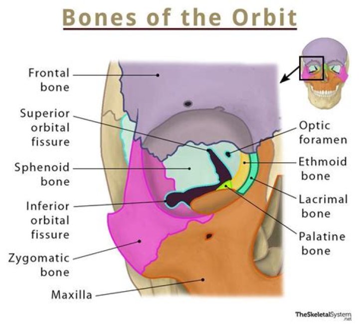
The orbital roof consists of two parts of two bones, the orbital plate frontal bone and the lesser wing of the sphenoid bone. … The medial orbital wall consists of four bones, the frontal process of the maxillary bone: the lacrimal bone, the orbital plate of the ethmoid bone, and the lesser wing of the sphenoid bone.
How many orbital bones are there?
The orbit, which protects, supports, and maximizes the function of the eye, is shaped like a quadrilateral pyramid, with its base in plane with the orbital rim. Seven bones conjoin to form the orbital structure, as shown in the image below.
What are the six major bones that form the orbit?
- Sphenoid.
- Frontal.
- Zygomatic.
- Ethmoid.
- Lacrimal.
- Maxilla.
- Palatine.
Where are the orbital bones?
By definition, the orbit (bony orbit or orbital cavity) is a skeletal cavity comprised of seven bones situated within the skull. The cavity surrounds and provides mechanical protection for the eye and soft tissue structures related to it.How many frontal bones are there?
Frontal boneArticulationsTwelve bones: the sphenoid, the ethmoid, the two parietals, the two nasals, the two maxillæ, the two lacrimals, and the two zygomatics
Where is maxillary?
The maxilla is the bone that forms your upper jaw. The right and left halves of the maxilla are irregularly shaped bones that fuse together in the middle of the skull, below the nose, in an area known as the intermaxillary suture.
What is an eye orbital?
The orbit is the bony cavity in the skull that houses the globe of the eye (eyeball), the muscles that move the eye (the extraocular muscles), the lacrimal gland, and the blood vessels and nerves required to supply these structures.
Is the orbital bone part of the skull?
The sphenoid and ethmoid bones located to the front of the skull form parts of the orbital sockets and nasal cavity; they also support and protect key organs found in the skull.How do you remember the orbital bones?
- My Little Eye Sits (in the orbit); or.
- Medial Layer Eye Socket.
The zygomatic bone (or zygoma) is a paired, irregular bone that defines the anterior and lateral portions of the face. The zygomatic complex is involved in the protection of the contents of the orbit and the contour of the face and cheeks.[1]
Article first time published onWhat is vomer bone?
The vomer is a small, thin, plow-shaped, midline bone that occupies and divides the nasal cavity. It articulates inferiorly on the midline with the maxillae and the palatines, superiorly with the sphenoid via its wings, and anterosuperiorly with the ethmoid.
What are the 14 facial bone?
The names of the 14 facial bones are: inferior nasal concha (2 of them,) lacrimal bones (2), mandible, maxilla (2), nasal bones (2), palatine bones (2), vomer, and zygomatic bones, or zygoma (2).
What is cheek bone?
zygomatic bone, also called cheekbone, or malar bone, diamond-shaped bone below and lateral to the orbit, or eye socket, at the widest part of the cheek. … It forms the central part of the zygomatic arch by its attachments to the maxilla in front and to the zygomatic process of the temporal bone at the side.
What are the 8 appendicular bones?
- Upper Limb.
- Shoulder girdle: Clavicle. Scapula. Arm. Humerus. Forearm. Radius. Ulna. Wrist or carpal bones. Scaphoid. Lunate. Triquetrum. Pisiform. Trapezium. …
- Lower Limb.
- Pelvic girdle (hip or coxal bone) Ilium. Ischium. Pubis. Thigh. Femur. Leg. Tibia. Fibula. Tarsal bones. Talas. Calcaneus. Cuboid.
What are orbits in the brain?
What is an Orbit? An orbit refers to the boney cavity occupied by your eye, nerves, muscles, fat, and additional soft tissues needed for proper eye movement and function. They are symmetrical and separated by the nasal cavity and paranasal sinuses. An orbit MRI scan is sometimes also referred to as an orbital MRI scan.
Where is the orbit of the earth?
Earth orbits the Sun at an average distance of 149.60 million km (92.96 million mi) in a counterclockwise pattern viewed above the northern hemisphere.
Which bones Protect eyes?
Parietal bones form the roof of the cranium and curve down to form the sides of the cranium. Also forming the sides of the cranium are the two temporal bones, located behind the eyes.
What is the palatine bone?
The Adult Palatine. The palatine bones contribute to the posterior part of the roof of the mouth and floor and lateral walls of the nose, the medial wall of the maxillary sinuses and the orbital floors. Each bone (Fig. 5-66) consists of horizontal and perpendicular plates (laminae) set at right angles to each other.
What is the jawbone called?
The lower jaw (mandible) supports the bottom row of teeth and gives shape to the lower face and chin. This is the bone that moves as the mouth opens and closes. The upper jaw (maxilla) holds the upper teeth, shapes the middle of the face, and supports the nose.
Where is sphenoid bone?
The sphenoid is an unpaired bone. It sits anteriorly in the cranium, and contributes to the middle cranial fossa, the lateral wall of the skull, and the floor and sides of both orbits. It has articulations with twelve other bones: Unpaired bones – Occipital, vomer, ethmoid and frontal bones.
Where is optic foramen?
The optic foramen, the opening through which the optic nerve runs back into the brain and the large ophthalmic artery enters the orbit, is at the nasal side of the apex; the superior orbital fissure is a larger hole through which pass large veins and nerves.…
What is a ethmoid bone?
The ethmoid bone is an unpaired cranial bone that is a significant component of the upper nasal cavity and the nasal septum. The ethmoid bone also constitutes the medial orbit wall.
What is a supraorbital foramen?
The supraorbital foramen or notch is the small opening at the central edge of the superior orbital margin in the frontal bone just below the superciliary arches that transmits the supra-orbital nerve, artery and vein.
What are the 22 bones of the skull?
The skull (22 bones) is divisible into two parts: (1) the cranium, which lodges and protects the brain, consists of eight bones (Occipital, Two Parietals, Frontal, Two Temporals, Sphenoidal, Ethmoidal) and the skeleton of the face, of fourteen (Two Nasals, Two Maxillae, Two Lacrimals, Two Zygomatics, Two Palatines, Two …
Which bone is not part of the eye orbit?
The maxilla is the only listed bone that is not part of the cranium. Instead, it is a facial bone. Which of the following bones is NOT part of the orbit? The temporal bone is lateral and too far posterior to contribute to the orbit.
What is in the temporal bone?
The temporal bone consists of a pair of bones that help make up the skull. Many cranial nerves and blood vessels pass through the temporal bone. Injuries to this bone can cause a loss of function in the facial muscles, as well as hearing loss and heavy bleeding.
What is malar area?
1. malar – the arch of bone beneath the eye that forms the prominence of the cheek. cheekbone, jugal bone, malar bone, os zygomaticum, zygomatic, zygomatic bone. jugal point, jugale – the craniometric point at the union of the frontal and temporal processes of the zygomatic bone.
What bones make lower jaw?
The mandible is the largest bone in the human skull. It holds the lower teeth in place, it assists in mastication and forms the lower jawline. The mandible is composed of the body and the ramus and is located inferior to the maxilla. The body is a horizontally curved portion that creates the lower jawline.
What is the maxillary crest?
This strip of bone is called the maxillary crest; it articulates in front with the quadrangular cartilage, and at the back with the vomer. The maxillary crest is described in the anatomy of the nasal septum as having a maxillary component and a palatine component.
Is the mandible a cranial bone?
The skull is composed of two parts: the cranium and the mandible. In humans, these two parts are the neurocranium and the viscerocranium (facial skeleton) that includes the mandible as its largest bone.
Is the palatine bone a cranial bone?
Primarily, the palatine bone serves a structural function, with its shape helping carve out important structures within the head and defining the lower wall of the inside of cranium. This bone helps form the nasal and oral cavities, the roof of the mouth, and the lower portion of the eye sockets (orbits).