What are the three layers of the neural tunic of the eye wall
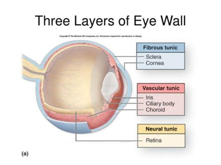
The eye is made up of three layers: the outer layer called the fibrous tunic, which consists of the sclera and the cornea; the middle layer responsible for nourishment, called the vascular tunic, which consists of the iris, the choroid, and the ciliary body; and the inner layer of photoreceptors and neurons called the …
What are the 3 layers tunics of the eye?
The eyeball has three layers: the outer fibrous tunic, the middle vascular tunic, and the inner sensory tunic.
What are the three layers or the neural tunic of the eye wall quizlet?
The eye is made up of three layers: the outer layer called the fibrous tunic, which consists of the sclera and the cornea; the middle layer responsible for nourishment, called the vascular tunic, which consists of the iris, the choroid, and the ciliary body; and the inner layer of photoreceptors and neurons called the …
What are the neural components of the eye?
Neural components: Include the retina and the optic nerve: Retina – cup-shaped outgrowth of the brain which forms the inner layer of the eyeball. Optic nerve – CNII responsible for vision.What are the three layers of the eye quizlet?
What are the three layers of the eye? The sclera, the choroid layer, and the retina.
What part of the eye consists of a pigmented layer and a neural layer?
The third and innermost layer of the eyeball is the retina. It consists of a pigmented layer and a neural layer. The pigmented layer is a sheet of melanin-containing epithelial cells that is located between the choroid and the neural part of the retina.
Which layer of the eye is considered the vascular layer?
The middle layer of the eye is the vascular tunic, which is mostly composed of the choroid, ciliary body, and iris. The choroid is a layer of highly vascularized connective tissue that provides a blood supply to the eyeball.
What are retinas?
The retina is a layer of tissue in the back of your eye that senses light and sends images to your brain. In the center of this nerve tissue is the macula. It provides the sharp, central vision needed for reading, driving and seeing fine detail. Retinal disorders affect this vital tissue.What is the inner layer of the retina?
Name of the layersContentLayer of PhotoreceptorInner and Outer segments of Photoreceptor
What layer of the eye is superficial and avascular?Cornea and sclera constitute the outer covering or coat of the eyeball. The main purpose of this coat is to protect structures inside the eye. The cornea is a transparent avascular tissue that acts as a structural barrier and protects the eye against infections.
Article first time published onWhat is the outer layer of the eye quizlet?
The outermost tissue layer of the eye includes the sclera, and the cornea. The part of the fibrous tunic that is embedded in the eye socket is a thick, opaque, white layer called the sclera.
How many layers does the middle tunic have quizlet?
The tunica media consists of 40-70 layers of elastic sheets alternating with smooth muscle.
What makes up the vascular tunic?
The vascular tunic is comprised of three distinct regions, (1) the iris, (2) the ciliary body, and (3) the choroid. The vascular tunic is mesodermal in origin and is situated between the outer fibrous tunic and the inner nervous tunic. The vascular tunic is also refered to as the uvea.
What are the layers of the eye in order from superficial to deep?
From deep to superficial, they are the inner limiting membrane, nerve fiber layer, ganglion cell layer, inner plexiform layer, inner nuclear layer, outer plexiform layer, outer nuclear layer, external limiting membrane, and the retinal pigment epithelium.
What are the two parts of the first tunic?
FIG. 869– Horizontal section of the eyeball. From without inward the three tunics are: (1) A fibrous tunic, (Fig. 869) consisting of the sclera behind and the cornea in front; (2) a vascular pigmented tunic, comprising, from behind forward, the choroid, ciliary body, and iris; and (3) a nervous tunic, the retina.
Which structures are a part of each tunic?
Tunics of the Eye: The outer coat, the fibrous tunica, of the eye includes the sclera, the white of the eye, and the cornea. The middle coat is called the uvea or the vascular tunic. This region includes the blood supply for the tunics of the eye.
Which layer of the eye is indicated by letter A?
Which layer of the eye is indicated by letter A? The iris, the visible colored part of the eye, is the most anterior portion of the vascular layer of the eye, and either dilates or constricts to control how much light enters the posterior chamber.
Which layer of the eye contains rods and cones?
Retina: a light sensitive layer that lines the interior of the eye. It is composed of light sensitive cells known as rods and cones.
What divides the eye into anterior and posterior?
The iris divides the eye into the anterior and posterior segments. The pupil can adjust its size independent of the iris.
What forms most of the pigmented vascular layer?
The angle formed by the iris and cornea contains connective tissue with endothelial channels called the trabecular meshwork, which drains aqueous humor in the anterior chamber into the venous canal of Schlemm[8]. From here, fluid drains into episcleral veins.
What is fibrous tunic?
The fibrous tunic is composed of the sclera and the cornea. The sclera covers nearly the entire surface of the eyeball. With its external surface being white-coloured, it is commonly known as the “white of the eye”. … The transparent cornea occupies the front center part of the external tunic.
How many layers are there in the eye?
The eye has three main layers. These layers lie flat against each other and form the eyeball. The outer layer of the eyeball is a tough, white, opaque membrane called the sclera (the white of the eye). The slight bulge in the sclera at the front of the eye is a clear, thin, dome-shaped tissue called the cornea.
What is outer nuclear layer?
The outer nuclear layer contains the cell bodies of the photoreceptor cells. These cells extend a process toward the outer plexiform layer, where they form the characteristic synaptic terminals of rods (spherules) and cones (pedicles).
What is ciliary epithelium?
The ciliary body is a part of the eye that includes the ciliary muscle, which controls the shape of the lens, and the ciliary epithelium, which produces the aqueous humor. The aqueous humor is produced in the non-pigmented portion of the ciliary body.
What is macula retina?
The macula is part of the retina at the back of the eye. It is only about 5mm across but is responsible for our central vision, most of our colour vision and the fine detail of what we see. The macula has a very high concentration of photoreceptor cells – the cells that detect light.
What are rods and cones?
Rods and cones are the receptors in the retina responsible for your sense of sight. They are the part of the eye responsible for converting the light that enters your eye into electrical signals that can be decoded by the vision-processing center of the brain. Cones are responsible for color vision.
What are the 3 functions of the cornea?
- to bend or refract light that enters the eye.
- to focus light entering the eye.
- to provide a physical barrier for the eye and protect it from germs, dust, etc.
Which tagged structure is part of the fibrous tunic?
Sclera. The sclera constitutes the major portion of the fibrous tunic and joins the cornea at the limbus. Anteriorly it is covered by bulbar conjunctiva beneath which is the episclera, the scleral stroma, and the lamina fusca.
What is the posterior portion of the middle layer of the eye?
The posterior segment or posterior cavity is the back two-thirds of the eye that includes the anterior hyaloid membrane and all of the optical structures behind it: the vitreous humor, retina, choroid, and optic nerve.
Is the translucent anterior section of the fibrous layer tunic of the eye?
The iris is the anterior portion of the vascular tunic and is visible through the cornea. The iris divides the anterior cavity of the eye into the anterior and posterior chambers.
What are the three color receptors?
In 1965 came experimental confirmation of a long expected result – there are three types of color-sensitive cones in the retina of the human eye, corresponding roughly to red, green, and blue sensitive detectors.