What does each wave of an ECG represent
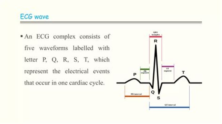
The different waves that comprise the ECG represent the sequence of depolarization and repolarization of the atria and ventricles. The ECG is recorded at a speed of 25 mm/sec (5 large squares/sec), and the voltages are calibrated so that 1 mV = 10 mm (2 large squares) in the vertical direction.
What does each wave of the ECG represent?
The different waves that comprise the ECG represent the sequence of depolarization and repolarization of the atria and ventricles. The ECG is recorded at a speed of 25 mm/sec (5 large squares/sec), and the voltages are calibrated so that 1 mV = 10 mm (2 large squares) in the vertical direction.
What do the P QRS and T waves represent?
The P wave in an ECG complex indicates atrial depolarization. The QRS is responsible for ventricular depolarization and the T wave is ventricular repolarization.
What are the 5 waves of an ECG?
- P wave. The P wave is a small deflection wave that represents atrial depolarization.
- PR interval. …
- QRS wave complex. …
- ST segment. …
- T wave. …
- Wave direction and size. …
- Interpreting the ECG. …
- Rate.
What does the R wave mean?
Image: R Wave. As you can see from the diagram, the R wave represents the electrical stimulus as it passes through the main portion of the ventricular walls. The wall of the ventricles are very thick due to the amount of work they have to do and, consequently, more voltage is required.
What is the Q wave on an ECG?
By definition, a Q wave on the electrocardiogram (ECG) is an initially negative deflection of the QRS complex. Technically, a Q wave indicates that the net direction of early ventricular depolarization (QRS) electrical forces projects toward the negative pole of the lead axis in question.
What does AP Wave represent?
The P wave represents the electrical depolarization of the atria. In a healthy person, this originates at the sinoatrial node (SA node) and disperses into both left and right atria.
What does V1 V2 V3 mean in ECG?
The areas represented on the ECG are summarized below: V1, V2 = RV. V3, V4 = septum. V5, V6 = L side of the heart. Lead I = L side of the heart.What does inverted T wave mean on ECG?
Inverted T waves. Ischemia: Myocardial ischemia is a common cause of inverted T waves. Inverted T waves are less specific than ST segment depression for ischemia, and do not in and of themselves convey a poor prognosis (as compared to patients with an acute coronary syndrome and ST segment depression).
What do tall R waves mean?Tall R waves in V1 can be caused by abnormal electrical conduction (RBBB or left-sided VT, which slowly spreads across the right ventricle, or a left-sided accessory pathway), loss of posterior myocardium (old or acute posterior MI) or chronic anterior hypertrophy (HCM), chronic or acute RV strain (RVH, PE), congenital …
Article first time published onWhat does enlarged R wave indicate?
The enlarged Q and R waves indicate myocardial infraction.
What does a flat P wave mean?
Absence of the P wave with a flat baseline may indicate: Fine atrial fibrillation. Sinoatrial arrest (with a secondary escape rhythm)
What is aq wave MI?
Q wave myocardial infarction refers to myocardial infarctions that in a Q wave forming on the 12-lead ECG once the infarction is completed.
What is ST and T wave abnormality?
“Primary” ST-T Wave Abnormalities (ST-T wave changes that are independent of changes in ventricular activation and that may be the result of global or segmental pathologic processes that affect ventricular repolarization): Drug effects (e.g., digoxin, quinidine, etc) Electrolyte abnormalities (e.g., hypokalemia)
What does ST and T wave abnormality mean?
Background: Nonspecific ST and T wave abnormalities (NSSTTA) on resting ECGs are associated with increased cardiovascular risk, and portend similar hazard ratios to traditional risk factors, such as dyslipidemia, hypertension, and diabetes mellitus (DM).
Are V1 V6 unipolar or bipolar?
The electrode leads each have a name. The bipolar extremity leads are called I, II and III. The unipolar extremity leads are called avR, avL and avF, and the chest leads are called V1–V6.
What does a wide P wave mean?
The presence of broad, notched (bifid) P waves in lead II is a sign of left atrial enlargement, classically due to mitral stenosis.
What rhythms have no P waves?
A junctional rhythm is characterized by QRS complexes of morphology identical to that of sinus rhythm without preceding P waves.
What is an abnormal P axis?
Abnormal P-wave axis is defined as any value outside 0–75° (Figure 1) (31). Figure 1. Representative ECG tracings of abnormal P-wave indices. A through (D), Prolonged P-wave duration (A), abnormal P-wave axis (B), abnormal P-wave terminal force in V1 (C), and advanced interatrial block (D).
What is a transmural Q wave infarction?
A transmural myocardial infarction refers to a myocardial infarction that involves the full thickness of the myocardium. It was one believed that the development of Q waves indicated the infarction was “transmural;” however, autopsy studies failed to confirm this.
When do Q waves appear after MI?
Q waves may develop within one to two hours of the onset of symptoms of acute myocardial infarction, though often they take 12 hours and occasionally up to 24 hours to appear.
What is a non Q wave MI?
Non–Q-wave myocardial infarction has been defined as acute myocardial infarction without a new-onset deep Q-wave on the ECG after day(s) of evolution, and because of the anatomopathological concept of infarction is usually related to necrosis, it results paradoxical to consider this widely known clinical and …
What does a low T wave mean?
T wave inversion less than 5 mm may still represents myocardial ischaemia, but is less severe than Wellens’ syndrome. Hypertrophic cardiomyopathy is the thickening of the left ventricle, occasionally right ventricle.
What is a normal T wave axis?
The frontal plane T-wave axis was estimated from 12-lead electrocardiograms obtained on admission and categorized as normal (15 degrees to 75 degrees ), borderline (75 degrees to 105 degrees or 15 degrees to -15 degrees ), and abnormal (>105 degrees or < -15 degrees ).
What does T wave changes mean?
T wave changes are secondary to electrolyte abnormalities in the myocardium since the ECG is representative of the electricity of the heart. The outflow of potassium from the myocyte during repolarization is necessary to restore resting membrane potential.