What is dual blood supply
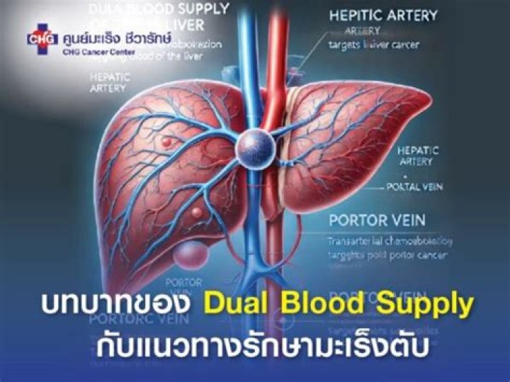
Liver dual arterial blood supply (LDABS) is a surgical procedure that shunts arterial blood to the portal vein system for enhancing liver blood supply to maintain liver regeneration.
What does it mean dual blood supply?
The liver receives a blood supply from two sources. The first is the hepatic artery which delivers oxygenated blood from the general circulation. The second is the hepatic portal vein delivering deoxygenated blood from the small intestine containing nutrients.
Why does the liver need a dual blood supply?
Your liver gets blood from two distinct sources: the hepatic artery and the portal vein. Oxygen-rich blood flows in through the hepatic artery, while nutrients from the intestines come through the portal vein. Remember the sinusoids? This is where they get all that oxygen- and nutrient-rich blood.
What is double blood supply for lung?
For the exchange of gases to occur effectively, the lungs require a dual vascular supply known as pulmonary and systemic circulations. The pulmonary circulation is responsible for bringing deoxygenated blood from the body to the lungs for gaseous exchange and then returning oxygenated blood to the heart.Which of the following has dual blood supply?
It is well known that the liver has a dual blood supply from the portal vein and hepatic artery. Approximately 75% of blood flow to the liver parenchyma is from the portal vein and 25% is from the hepatic artery; 50% of oxygenation is derived from the portal vein and hepatic artery each.
How is vascularization of the liver different from other organs?
The liver’s blood supplies Every two and a half minutes a gallon of blood passes through the liver’s complicated network of arteries, veins and capillaries. Unlike any other organ in the body, the liver has two blood supplies: 75% of its blood comes in through the portal vein system.
What is Hepatoportal system?
The hepatic portal system is the venous system that returns blood from the digestive tract and spleen to the liver (where raw nutrients in blood are processed before the blood returns to the heart).
What is a Lingula?
The term lingula refers to the tip or tongue-like projection of the upper lobe of the left lung but in general it is considered also to be the entire portion of this segment which is supplied by the first segmental bronchus that arises from the upper lobe bronchus.What is blood supply of lungs?
The bronchial artery is the main source of blood supply for the bronchi. The bronchial arteries provide the normal systemic arterial supply to the lungs after birth and generally comprise one to three small arteries from the upper dorsal aorta or right intercostal artery supplying each lung (,6),(,10).
Why is my left lung bigger?A person’s lungs are not the same size. The right lung is a little wider than the left lung, but it is also shorter. According to York University, the right lung is shorter because it has to make room for the liver, which is right beneath it. The left lung is narrower because it must make room for the heart.
Article first time published onWhat are the 5 functions of the liver?
- Bile production and excretion.
- Excretion of bilirubin, cholesterol, hormones, and drugs.
- Metabolism of fats, proteins, and carbohydrates.
- Enzyme activation.
- Storage of glycogen, vitamins, and minerals.
- Synthesis of plasma proteins, such as albumin, and clotting factors.
What is the name given to liver cells?
Liver cells, or hepatocytes, have direct access to the liver’s blood supply through small capillaries called sinusoids. Hepatocytes carry out many metabolic functions, including the production of bile.
Where does the blood go after the liver?
Blood is carried out of the liver through the hepatic veins to the heart.
What is the longest vein in the body?
Great Saphenous Vein (GSV) – The GSV is the large superficial vein of the leg and the longest vein in the entire body. It can be found along the length of the lower limb, returning blood from the thigh, calf, and foot to the deep femoral vein at the femoral triangle. The femoral triangle is located in the upper thigh.
What is in the circle of Willis?
The Circle of Willis is the joining area of several arteries at the bottom (inferior) side of the brain. At the Circle of Willis, the internal carotid arteries branch into smaller arteries that supply oxygenated blood to over 80% of the cerebrum.
What delivers blood to the kidney?
Blood comes to the kidneys from the abdominal aorta and inferior vena cava, the large arteries and veins that are part of the ascending aorta. Oxygenated blood is brought to the kidneys from a small branch called the renal artery.
What are the 3 portal systems?
Examples of such systems include the hepatic portal system, the hypophyseal portal system, and (in non-mammals) the renal portal system. Unqualified, portal venous system often refers to the hepatic portal system. For this reason, portal vein most commonly refers to the hepatic portal vein.
What is portal circulation class 11?
Hepatic Portal System There is special vascular connection that exists between the digestive tract and liver in all chordates and is called as hepatic portal system. This system carries blood from intestine to the liver before it is delivered to the systemic circulation.
What organs does the hepatic portal vein drain?
The portal vein or hepatic portal vein (HPV) is a blood vessel that carries blood from the gastrointestinal tract, gallbladder, pancreas and spleen to the liver.
Which organ receives oxygenated and deoxygenated?
Spleen is situated in the upper far left part of the abdomen and primarily acts as a blood filter. It receives oxygenated blood only while lungs and gills receive deoxygenated blood.
How do hepatocytes detoxify?
Toxins enter hepatocytes via a dual blood supply provided by the hepatic artery and portal vein, where they encounter a wide variety of high-volume biochemical reactions that collectively facilitate removal of these chemicals from the body.
Does the liver produce insulin?
The liver both stores and produces sugar… The need to store or release glucose is primarily signaled by the hormones insulin and glucagon. During a meal, your liver will store sugar, or glucose, as glycogen for a later time when your body needs it.
How does blood supply to the heart?
Coronary arteries supply blood to the heart muscle. Like all other tissues in the body, the heart muscle needs oxygen-rich blood to function. Also, oxygen-depleted blood must be carried away. The coronary arteries wrap around the outside of the heart.
What are the three main arteries that supply blood to the digestive organs?
The major arteries supplying the gastrointestinal tract are the celiac, superior mesenteric, and inferior mesenteric arteries.
How many secondary bronchi are in the right lung?
The right main bronchus subdivides into three secondary bronchi (also known as lobar bronchi), which deliver oxygen to the three lobes of the right lung—the superior, middle and inferior lobe.
What is cardiac notch?
It is the cardiac notch is the lateral deflection of the anterior border of the left lung. It is produced to accommodate the space taken up by the heart.
What is the tongue of the lung?
The lingula of the lung is a tongue-shaped region of the left lung. It is also known by its Latin name, lingula pulmonis sinistri, which means little tongue of the left lung.
What is the horizontal fissure?
The horizontal fissure (also called the minor fissure) is a unilateral structure in the right lung that separates the right middle lobe from the right upper lobe.
How far do lungs go down back?
The lungs are found in the chest on the right and left side. At the front they extend from just above the collarbone (clavicle) at the top of the chest to about the sixth rib down. At the back of the chest the lungs finish around the tenth rib.
What organ causes left lung?
The right and left lungs differ in size and shape to accommodate other organs that encroach on the thoracic region. The right lung consists of three lobes and is shorter than the left lung, due to the position of the liver underneath it. The left lung consist of two lobes and is longer and narrower than the right lung.
Which lung is upper?
The right upper lobe of the lung is located in the right superior corner of the thoracic cavity lateral to the trachea and esophagus. It is superior to the horizontal and oblique fissures, which separates the upper lobe from the middle and lower lobes of the right lung.