What is intercostal artery
The intercostal arteries are a group of arteries that supply the area between the ribs (“costae”), called the intercostal space.
Where does the intercostal artery come from?
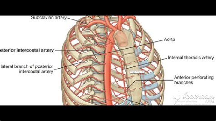
The first and second posterior intercostal arteries originate from the superior (supreme) intercostal artery, a branch of the costocervical trunk. Third to eleventh posterior intercostal arteries arise directly from the posterior surface of the thoracic aorta.
Where is the intercostal vein located?
The intercostal veins are a group of veins which drain the area between the ribs (“costae”), called the intercostal space. Posterior intercost veins that drain into the Superior intercostal vein – 2nd, 3rd, and 4th intercostal spaces. The superior intercostal vein then drains into the Azygous vein.
What is the superior intercostal artery?
The supreme intercostal arteries, or superior intercostal arteries, are formed as a direct result of the embryological development of the intersegmental arteries. These arteries are paired structures of the upper thorax which normally form to provide blood flow to the first and second posterior intercostal arteries.What is posterior intercostal arteries?
The posterior intercostal arteries are branches of the superior intercostal artery (upper two spaces) and the descending aorta (lower nine spaces). They supply the chest wall, parietal pleura, and, through their dorsal branches, the skin and muscles of the back and the spine and its contents.
What supplies the posterior chest wall?
Three arteries supply each intercostal space; the posterior intercostal artery and two branches of anterior intercostal arteries. These intercostal blood vessels run along with the nerves between the internal intercostal muscle and innermost intercostal muscles in the costal groove.
What supplies anterior intercostal arteries?
The musculophrenic artery supplies the anterior intercostal arteries of spaces 7 to 9 after it branches off the internal thoracic.
What are the branches of subclavian artery?
Branches. The subclavian arteries give off five major arteries each: the vertebral artery, the internal thoracic artery, the thyrocervical trunk, the costocervical trunk, and the dorsal scapular artery.Where do the intercostal veins drain?
The intercostal veins run above the intercostal arteries, which run above the intercostal nerves. The posterior intercostal veins drain into the azygos vein (right side), and hemiazygos and accessory hemiazygos veins (left side). The latter two veins ultimately drain into the azygos vein.
Which posterior intercostal arteries are longer the right or the left?The posterior intercostal arteries arise from the back of the thoracic aorta. On the right side, these arteries are longer because the aorta lies to the left of the midline. They travel in front of the vertebrae and behind vessels of the azygos venous system, esophagus, and thoracic duct.
Article first time published onHow many intercostal veins do we have?
There are eleven posterior intercostal veins on each side. Their patterns are variable, but they are commonly arranged as: The 1st posterior intercostal vein, supreme intercostal vein, drains into the brachiocephalic vein or the vertebral vein.
Where is the first intercostal space?
Intercostal spaceTA21102FMA12243Anatomical terminology
What vein drains the first intercostal space?
One posterior and two anterior intercostal veins occupy each intercostal space. Anteriorly, they drain into the musculophrenic and internal thoracic veins. Posterior venous drainage is more anatomically variable. The first posterior intercostal veins drain into the vertebral vein or the brachiocephalic vein.
How many posterior intercostal arteries are there?
Gross Anatomy. There are 11 paired arteries that constitute the posterior intercostal arteries. The first two intercostal spaces are supplied by the superior intercostal artery, and the remaining nine are supplied by separate branches from the descending thoracic aorta 1.
What is inferior chest?
Inferiorly, its extent is defined by the lower border of the axillary fossa. The anterior border includes the pectoralis muscles, and the posterior border includes the latissimus dorsi, which are both visible at the skin surface as the anterior and posterior axillary folds, respectively.
What's the main artery called?
The largest artery is the aorta, the main high-pressure pipeline connected to the heart’s left ventricle. The aorta branches into a network of smaller arteries that extend throughout the body. The arteries’ smaller branches are called arterioles and capillaries.
Is chest wall the ribs?
The skin, fat, muscles, bones, and other tissues that form a protective structure around vital organs in the area between the neck and the abdomen, including the heart, major blood vessels, lungs, and liver. The bones in the chest wall include the ribs, sternum (breastbone), and spine.
What are intercostal nerves?
The intercostal nerves emerge from the somatic nervous system and aid in the contraction of muscles as well as provide sensory information from the skin and parietal pleura. The intercostal nerves arise from the anterior rami of the thoracic spinal nerves from T1 to T11.
What vein does the posterior intercostal vein drain to?
One posterior and two anterior intercostal veins occupy each intercostal space. Anteriorly, they drain into the musculophrenic and internal thoracic veins. Posterior venous drainage is more anatomically variable. The first posterior intercostal veins drain into the vertebral vein or the brachiocephalic vein.
What is the left superior intercostal vein?
The left superior intercostal vein forms by the union of the 2nd to 4th left posterior intercostal veins. It courses superiorly to the left of the midline, arches posteriorly lateral to the aortic arch to drain into the left brachiocephalic vein. It typically communicates with the accessory hemiazygos vein.
What is the purpose of subclavian artery?
The subclavian arteries lie just below the clavicles, providing blood supply to the bilateral upper extremities with contributions to the head and neck. The right subclavian artery derives from the brachiocephalic trunk, while the left subclavian artery originates directly from the aortic arch.
Is subclavian a vein?
The subclavian vein (SVC) is classified as a deep vein and is the major venous channel that drains the upper extremities. Other deep veins of the upper extremity that accompany the major arteries include the radial, ulnar, brachial, axillary veins.
What is the first branch of the subclavian artery?
The branches of the subclavian artery are the vertebral artery, the internal mammary (thoracic) artery, the thyrocervical trunk and the costocervical trunk (Fig. 7.7). The vertebral artery is the first branch of the subclavian artery.
Where does the subclavian artery become the axillary artery?
The subclavian arteries course laterally between the anterior and middle scalene muscles. The distal limit of the subclavian artery is the lateral border of the first rib, where it becomes the axillary artery.
How many intercostal anterior spaces are there?
The intercostal spaces, also known as interspaces, are the space between the ribs. There are 11 spaces on each side and they are numbered according to the rib which is the superior border of the space.
What does the subclavian vein drain?
The primary function of the subclavian vein is to drain deoxygenated blood from the upper region of the body—including the arms and the shoulder areas—and transport it back to the heart. 6 Another important function of the subclavian is to collect lymph fluid from the lymphatic system from the internal jugular vein.
What drains blood from the head and neck?
jugular vein, any of several veins of the neck that drain blood from the brain, face, and neck, returning it to the heart via the superior vena cava. The main vessels are the external jugular vein and the interior jugular vein.
Where is the 8th rib?
In the anterior thorax, the first 7 pairs of ribs are attached to the sternum or breastbone by cartilage. The lower 5 ribs do not attach to the sternum. The 8th, 9th, and 10th ribs are attached to each other by costal cartilage.
How many ribs do human have?
In humans there are normally 12 pairs of ribs. The first seven pairs are attached directly to the sternum by costal cartilages and are called true ribs.
What is the sixth intercostal space?
Locate the 6th intercostal space by counting parasternally from the clavicle or from the manubriosternal synchondrosis (→ 3.5). Follow the course of the intercostal space in a lateral direction and locate SP-21 on the axillary midline (note: the intercostal space laterally curves upward).
What is the longest vein in the body?
Great Saphenous Vein (GSV) – The GSV is the large superficial vein of the leg and the longest vein in the entire body. It can be found along the length of the lower limb, returning blood from the thigh, calf, and foot to the deep femoral vein at the femoral triangle. The femoral triangle is located in the upper thigh.