What is the H test for eyes
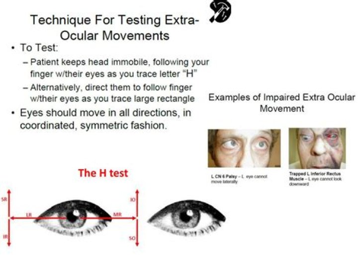
The test itself is simple. Your eye healthcare provider or technician will ask you to sit up straight while you stare at an object in front of you, which is usually a pen, fixation light, or small picture held 12 and 16 inches away. They will move the object up and down and side to side in an H-shaped pattern.
What does the Double H test examine?
Slow tracking, or “pursuits” are assessed by the ‘follow my finger’ test, in which the examiner’s finger traces an imaginary “double-H”, which touches upon the eight fields of gaze. These test the inferior, superior, lateral and medial rectus muscles of the eye, as well as the superior and inferior oblique muscles.
What does the six cardinal fields of gaze test for?
The corneal light reflex, cover/uncover test, and six cardinal fields of gaze are used to assess extraocular muscle function. The corneal light reflex may be used to assess extraocular muscle imbalance. The cover/uncover test is used to determine misalignment of the eye.
What is eye motility?
The term ocular motility refers to the study of the twelve extraocular muscles and their impact on eye movement. Each eye has six muscles, four rectus and two oblique, which, when functioning properly, allow the eyes to work together in a wide range of gaze. Muscles of the Right Eye.What is EOM in eye?
EOM; Extraocular movement; Ocular motility examination. Extraocular muscle function testing examines the function of the eye muscles. A health care provider observes the movement of the eyes in eight specific directions. The eye is the organ of sight, a nearly spherical hollow globe filled with fluids (humors).
What are the symptoms of weak eye muscles?
- Sore, tired, burning or itching eyes.
- Watery or dry eyes.
- Blurred or double vision.
- Headache.
- Sore neck, shoulders or back.
- Increased sensitivity to light.
- Difficulty concentrating.
- Feeling that you cannot keep your eyes open.
What is the test for peripheral vision?
The visual field test is a subjective measure of central and peripheral vision, or “side vision,” and is used by your doctor to diagnose, determine the severity of, and monitor your glaucoma. The most common visual field test uses a light spot that is repeatedly presented in different areas of your peripheral vision.
How do I check my ophthalmology motility?
When testing motility, assess the eye alignment in primary gaze (consider using the Hirschberg test) and then have the patient move the eyes in an “H” pattern, as shown in Figure 1. Using your finger, a light or a toy, trace an “H” pattern in front of the patient while instructing them to hold their head still.Can Vitamin B12 deficiency cause nystagmus?
Although vitamin B12 deficiency can cause eye movement disorders (1), including downbeat nystagmus (1-3), upbeat nystagmus in vitamin B12 deficiency has not been previously reported.
What are the 6 ocular movements?You are now familiar with the 6 cardinal directions of gaze (right/up; right; right/down; left/up; left; left/down), as well as the remainder of the yoked eye movements (straight up; straight down; convergence).
Article first time published onWhat produces conjunctiva?
The conjunctiva helps lubricate the eye by producing mucus and tears, although a smaller volume of tears than the lacrimal gland. It also contributes to immune surveillance and helps to prevent the entrance of microbes into the eye.
Can I drive if I fail a field vision test?
Field of Vision Tests If they deem that your peripheral vision is not adequate enough, they revoke your driving licence. From a legal stance, this would make it illegal to continue driving, as it is an offence to drive without a valid driving licence.
What is the normal range of peripheral vision?
This type of vision is the result of different nerve cells and rods located outside of the macula. As compared to animals, humans have a limited peripheral view. A normal visual field for a person covers 170 degrees around, while peripheral vision covers 100 degrees of this field.
Can peripheral vision be corrected?
In many cases of PVL, your side vision may not be restored. It’s important to see an eye doctor regularly to monitor and diagnose conditions that may affect your PVL permanently. Your doctor may be able to suggest certain lifestyle changes you can make if you have PVL.
Can neck problems affect your eyes?
Muscle tension in the upper back, neck and shoulders can lead to headaches or problems with your vision, as the flow of blood is restricted to your eyes. Signs you may notice are: Throbbing pain around the temples. Blurred vision or difficulty focusing.
How can I make my eye nerves stronger?
- Eat Well. Good eye health starts with the food on your plate. …
- Quit Smoking. …
- Wear Sunglasses. …
- Use Safety Eyewear. …
- Look Away From the Computer Screen. …
- Visit Your Eye Doctor Regularly.
Can damaged eye muscles be repaired?
Eye muscle repair surgery helps realign the eyes so that both point in the same direction. This procedure is most often performed on children with strabismus, but it may also be done to help adults with eye muscle problems.
Can a vitamin deficiency cause nystagmus?
Together with the neuropsychiatric features usually associated with this condition, a downbeat nystagmus syndrome was observed. It is concluded that vitamin B12 deficiency may also result in lesions to those cerebellar or brain-stem structures that are generally assumed to cause downbeat nystagmus.
What are the symptoms of B12 deficiency?
- a pale yellow tinge to your skin.
- a sore and red tongue (glossitis)
- mouth ulcers.
- pins and needles (paraesthesia)
- changes in the way that you walk and move around.
- disturbed vision.
- irritability.
- depression.
What is thiamine for prescribed?
Thiamine is used to treat beriberi (tingling and numbness in feet and hands, muscle loss, and poor reflexes caused by a lack of thiamine in the diet) and to treat and prevent Wernicke-Korsakoff syndrome (tingling and numbness in hands and feet, memory loss, confusion caused by a lack of thiamine in the diet).
Which of the following symptoms would you expect a person suffering from Abducens nerve paralysis to display?
Patients who develop abducens nerve palsy often present with binocular horizontal diplopia, which is double vision when looking at objects side by side. There will be a notable weakness of the ipsilateral lateral rectus muscle leading to a deficit in eye abduction on the affected side.
Where is the ocular nerve?
Made of nerve cells, the optic nerve is located in the back of the eye. Also known as the second cranial nerve or cranial nerve II, it is the second of several pairs of cranial nerves.
What happens when the Trochlear nerve is damaged?
Patients with trochlear nerve palsy complain of double vision vertically (vertical diplopia) or the images being tilted or rotated (torsional diplopia). The diplopia is binocular and may worsen or improve in different gazes.
How many cardinal gazes are there?
The six cardinal positions of eye gaze isolate the individual extraocular muscles. In order to determine which muscle is abnormal, the six positions of gaze are observed to determine if the eye moves in each direction.
What is cardinal direction of gaze?
The “H” path provides the six cardinal positions of gaze, which we divide into horizontal and vertical gaze. A. Horizontal gaze comprises abduction (lateral rotation) and adduction (medial rotation). … The medial rectus produces eye adduction.
What cranial nerve is Cardinal gaze?
Cranial nerves III, IV, and VI—Oculomotor, trochlear, and abducens. Test extraocular movements in the six cardinal directions of gaze, and look for loss of conjugate movements in any direction.
How do you score NIH on intubated patient?
The intubated patient should be asked to write. The patient in a coma (item 1a=3) will automatically score 3 on this item. The examiner must choose a score for the patient with stupor or limited cooperation, but a score of 3 should be used only if the patient is mute and follows no one-step commands.
What is the difference between the sclera and conjunctiva?
The conjunctiva contributes to the tear film and protects the eye from foreign objects and infection. The sclera is the thick white sphere of dense connective tissue that encloses the eye and maintains its shape.
What does normal conjunctiva look like?
Normal: In a normal patient, the sclera is white in color and the palpebral conjunctiva appears pink. Unless conjunctiva is diseased you are only visualizing sclera and palpebral vascular bed through the translucent conjunctiva.
What is injected conjunctiva?
Injected conjunctiva. This is a red eye caused by dilation of blood vessels in the conjunctiva. It can have many causes. [Read more about red eyes.]