What is the remnant of the ductus arteriosus called
Ligamentum arteriosum (also known as Ligament of Botallo or Harvey’s ligament) is a fibrous remnant of the fetal ductus arteriosus (ductus Botalli, Botallo’s duct). The ductus arteriosus is a vessel connecting the pulmonary trunk and the aortic arch in the fetus.
Which of the following is a remnant of ductus arteriosus?
The adult remnant of the ductus arteriosus is called the ligamentum arteriosum. It is located between the pulmonary trunk and the aorta.
What is the ductus arteriosus called after birth?
It allows most of the blood from the right ventricle to bypass the fetus’s fluid-filled non-functioning lungs. Upon closure at birth, it becomes the ligamentum arteriosum.
What is the name of the remnant that remains after birth?
Structural closure in term babies occurs within 3 to 7 days. After it closes, the remnant is known as ligamentum venosum. If the ductus venosus fails to occlude after birth, it remains patent (open), and the individual is said to have a patent ductus venosus and thus an intrahepatic portosystemic shunt (PSS).Why is it called ligamentum arteriosum?
Arterial bunchFromleft pulmonary arteryTodescending aortaIdentifiersLatinLigamentum arteriosum
Where does the ductus arteriosus originate from?
The ductus arteriosus is a remnant of the distal sixth aortic arch and connects the pulmonary artery at the junction of the main pulmonary artery and the origin of the left pulmonary artery to the proximal descending aorta just after the origin of the left subclavian artery.
What does the ductus arteriosus do?
The ductus arteriosus is a normal blood vessel that connects two major arteries — the aorta and the pulmonary artery — that carry blood away from the heart. The lungs are not used while a fetus is in the womb because the baby gets oxygen directly from the mother’s placenta.
What causes ductus arteriosus to close?
The increased arterial oxygen tension and decrease in blood flow through the ductus arteriosus causes the ductus to constrict and functionally close by 12 to 24 hours of age in healthy, full-term newborns, with permanent (anatomic) closure occurring within 2 to 3 weeks.What happens to the foramen ovale ductus arteriosus and ductus venosus after birth?
Review of respiratory changes and other changes at birth. As soon as the baby is born, the foramen ovale, ductus arteriosus ductus venosus and umbilical vessels are no longer needed. The sphincter in the ductus venosus constricts, so that all blood entering the liver passes through the hepatic sinusoids.
What is it called when your placenta doesn't come out?When the placenta successfully detaches from the uterine wall but fails to be expelled from the woman’s body it is considered a trapped placenta. This usually happens as a result of the cervix closing before the placenta has been expelled. The Trapped Placenta is left inside the uterus.
Article first time published onWhat does the ductus arteriosus become?
The ductus arteriosus responds to these changes by closing and becoming the ligamentum arteriosum. This prevents oxygenated blood from returning to the pulmonary circulation and after passing through the lungs and into the aorta.
What is ductus Caroticus?
Ductus Caroticus was a portion of the embryonic. dorsal aorta between points of junction with the third and. fourth branchial or aortic arch arteries, usually, it disappears in. early embryonic development.
What does truncus arteriosus mean?
Truncus arteriosus is a birth defect of the heart. It occurs when the blood vessel coming out of the heart in the developing baby fails to separate completely during development, leaving a connection between the aorta and pulmonary artery.
Is ductus arteriosus same as ligamentum arteriosum?
Ligamentum arteriosum (also known as Ligament of Botallo or Harvey’s ligament) is a fibrous remnant of the fetal ductus arteriosus (ductus Botalli, Botallo’s duct). The ductus arteriosus is a vessel connecting the pulmonary trunk and the aortic arch in the fetus.
Is ligamentum arteriosum ductus arteriosus?
The ligamentum arteriosum (or arteriosus) is the small fibrous remnant of the fetal ductus arteriosum, located between and connecting the proximal left pulmonary artery and the undersurface of the junction of the aortic arch and descending aorta, at the aortic isthmus.
Where is the location of the ductus arteriosus?
Anatomy. In the normal heart with a left-sided aortic arch, the ductus arteriosus connects the left pulmonary artery near its origin to the descending aorta just distal to the left subclavian artery. The ductus arteriosus may persist in a wide variety of sizes and configurations.
What is the function of the ductus arteriosus quizlet?
In the developing fetus, the ductus arteriosus, is a blood vessel connecting the pulmonary artery to the aortic arch. It allows most of the blood from the right ventricle to bypass the fetus’s fluid-filled non-functioning lungs. Upon closure at birth, it becomes the ligamentum arteriosum.
Which statement below accurately describes the role of the ductus arteriosus?
The answer is C. This is the only correct statement about the ductus arteriosus. This structure connects the pulmonary artery and aorta, which helps carry mixed blood (oxygenated and deoxygenated blood) to the lower body and back to the placenta via the umbilical arteries (which branch off the descending aorta).
What is the role of ductus arteriosus in Bird's circulatory system?
During embryonic development, reptiles, birds, and mammals all possess either one or two paired ductus arteriosi that provide a fetal shunt of blood away from the lungs.
What is the function of the ductus arteriosus shunt?
This shunt moves blood from the right atrium of the heart to the left atrium. The ductus arteriosus moves blood from the pulmonary artery to the aorta. Oxygen and nutrients from the mother’s blood are sent across the placenta to the fetus.
What is the ductus arteriosus and which structures does it connect?
The ductus arteriosus is a vascular structure that connects the main pulmonary artery (MPA) with the descending aorta or the subclavian artery (SA).
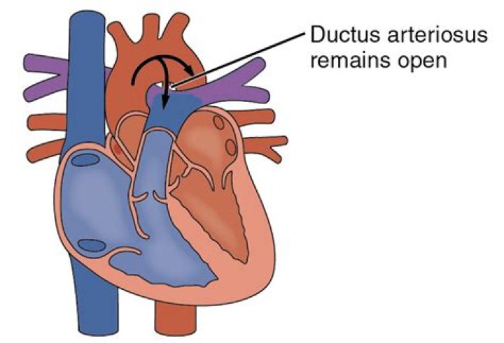
How does patent ductus arteriosus occur?
In PDA, abnormal blood flow occurs between two of the major arteries connected to the heart (the aorta and the pulmonary artery). This happens because a blood vessel called the ductus arteriosus doesn’t close after birth as it should.
What is the remnant of the umbilical artery?
The umbilical cord vein is the remnant of embryological venous development that results in the obliteration of the right umbilical cord vein and the establishment of two pathways through the liver and heart for oxygenated blood travelling from the placenta to the foetus via the persisting left umbilical cord vein.
What do the ductus arteriosus and the foramen ovale become at birth?
When do the ductus arteriosus and the foramen ovale become the adult structures? upon birth they change immediately into the ligamentum arteriosum and the fossa ovalis, respectively.
What is the remnant of the umbilical vein?
The ligamentum teres is the remnant of the umbilical vein working throughout fetal life. Initially a pair of the umbilical veins entered the sinus venosus.
What is persistent ductus arteriosus?
Patent ductus arteriosus (PDA) is a persistent opening between the two major blood vessels leading from the heart. The opening (ductus arteriosus) is a normal part of a baby’s circulatory system in the womb that usually closes shortly after birth. If it remains open, it’s called a patent ductus arteriosus.
How do you close ductus arteriosus?
In a catheter procedure, a thin tube (catheter) is inserted into a blood vessel in the groin and threaded up to the heart. Through the catheter, a plug or coil is inserted to close the ductus arteriosus. If the procedure is done on an outpatient basis, you or your child probably won’t stay overnight in the hospital.
What drug closes the ductus arteriosus?
Indomethacin is a non-steroidal anti-inflammatory drug that is a potent inhibitor of prostaglandin E(2) synthesis. After birth, the ductus arteriosus closes spontaneously within 2-4 days in term infants. The major factor closing the ductus arteriosus is the tension of oxygen, which increases significantly after birth.
Where do they cut for episiotomy?
An episiotomy is a cut (incision) through the area between your vaginal opening and your anus. This area is called the perineum. This procedure is done to make your vaginal opening larger for childbirth.
What do hospitals do with placenta after birth?
Hospitals treat placentas as medical waste or biohazard material. The newborn placenta is placed in a biohazard bag for storage. Some hospitals keep the placenta for a period of time in case the need arises to send it to pathology for further analysis.
What happens if you have a miscarriage and don't get cleaned out?
Often, some of the pregnancy tissue remains in the uterus after a miscarriage. If it is not removed by scraping the uterus with a curette (a spoon-shaped instrument), you may bleed for a long time or develop an infection.