What is the spring ligament
The plantar calcaneonavicular ligament also referred to as spring ligament is a thick wide band of cartilaginous connective tissue that supports the medial longitudinal arch of the foot, failure in the spring ligament leads to flat foot deformity.
Why is it called the spring ligament?
The plantar calcaneonavicular ligament, by supporting the head of the talus, is principally concerned in maintaining the arch of the foot. … This ligament contains a considerable amount of elastic fibers, so as to give elasticity to the arch and spring to the foot; hence it is sometimes called the “spring” ligament.
Can you tear your spring ligament?
A spring ligament tear can occur because of failure of the tibialis posterior tendon in adult-acquired flatfoot deformity or as an isolated injury with a normal tibialis posterior tendon. The superomedial spring ligament is the most common site of rupture.
What is the spring ligament ankle?
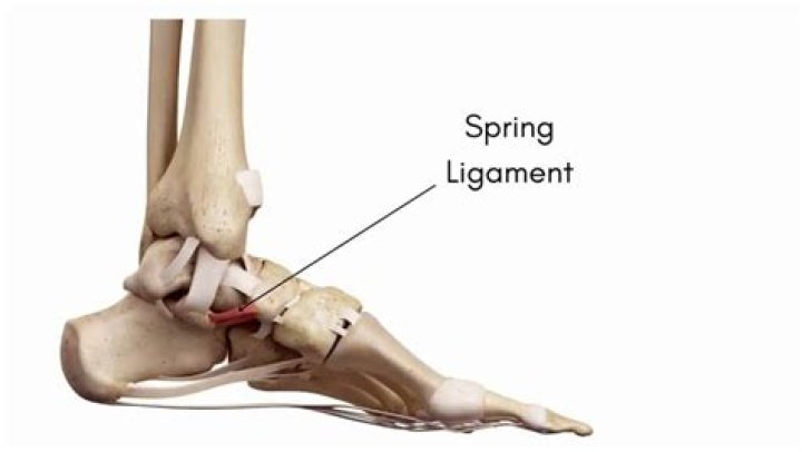
Spring Ligament. The plantar calcaneonavicular ligament (also known as the spring ligament) is a ligament on the underside of the foot that connects the calcaneus and the navicular bone.What is the spring ligament complex?
The spring ligament complex (SLC; also known as plantar calcaneonavicular ligament) is a group of ligaments that connect the sustentaculum talus of calcaneus to the plantar aspect of the navicular bone of the foot, supporting the talar head as part of the anterior talocalcaneonavicular joint [1].
How do you heal a spring ligament?
Reconstruction or augmentation methods for spring ligament tears or laxity include the use of anterior deltoid, peroneal tendon graft, split thickness tibialis anterior tendon graft, posterior tibial tendon stump, or suture repair.
Is the spring ligament part of the deltoid?
The superficial deltoid originates from the anterior & inferior aspects of medial malleolus fanning out & sending 3 bands to navicular and along plantar calcaneonavicular (spring) ligament, to sustenaculum tali of calcaneus and to medial tubercle. It is also partially covered by tendon sheaths & crural fascia.
What causes posterior tibial tendonitis?
What causes Posterior Tibial Tendonitis? Posterior tibial tendon dysfunction often happens due to repetitive overuse. Dancers and athletes who play high impact sports are at risk due to the stress they place on this tendon. An acute injury, such as a fall or collision, can also tear the posterior tibial tendon.Can you walk with a torn ligament in your foot?
The quick answer is yes, typically you can walk with a torn ligament or tendon in the foot. Walking may be painful but you can typically still walk. For example, the Posterior Tibialis Tendon runs down the back of the shin, behind the middle bump of the ankle (medial malleolus) and to the bottom of the foot.
What ligament is usually torn in a Grade 1 inversion ankle sprain?If you have suffered an inversion ankle sprain it means you have injured one or more of the three main ligaments on the outside of your ankle; the anterior talofibular ligament (ATFL), the posterior talofibular ligament (PTFL) and the calcaneofibular ligament (CFL).
Article first time published onWhat is the plantar ligament?
Plantar means ‘foot’ and fascia means ‘band’. Thus, the plantar fascia ligament is a thickened fibrous aponeurosis that originates from the medial tubercle of the calcaneus (heel bone) inserts on the metatarsal heads (ball of the foot) and then forms the fibrous flexor sheaths on the plantar aspect of the toes.
Where is the Lisfranc ligament?
The Lisfranc ligament is a large band of plantar collagenous tissue that spans the articulation of the medial cuneiform and the second metatarsal base.
Where is the posterior Talofibular ligament?
Posterior talofibular ligament. The posterior talofibular ligament originates from the malleolar fossa, located on the medial surface of the lateral malleolus, coursing almost horizontally to insert in the posterolateral talus.
What is Talofibular ligament?
It is one of the lateral ligaments of the ankle and prevents the foot from sliding forward in relation to the shin. … It is the most commonly injured ligament in a sprained ankle—from an inversion injury—and will allow a positive anterior drawer test of the ankle if completely torn.
What is bifurcate ligament?
The bifurcated ligament (internal calcaneocuboid, interosseous ligament or bifurcate ligament) is a strong band, attached behind to the deep hollow on the upper surface of the calcaneus and dividing in front in a Y-shaped manner into a calcaneocuboid and a calcaneonavicular part.
What is the ankle joint in medical terms known as?
The ankle joint (or talocrural joint) is a synovial joint located in the lower limb. It is formed by the bones of the leg (tibia and fibula) and the foot (talus).
What are the 4 ligaments of the deltoid ligament?
The deltoid ligament is composed of 4 fičera: 1. Anterior tibiotalar ligament 2. Tibiocalcaneal ligament 3. Posterior tibiotalar ligament 4.
What is the deltoid ligament?
The deltoid ligament, also known as the medial collateral ligament complex, is a strong, broad ligament with multifascicular appearance that spans out from the medial malleolus toward the talus, calcaneus, and navicular bones.
What are the 3 bones that make up the ankle?
- The shin bone (tibia)
- The thinner bone running next to the shin bone (fibula)
- A foot bone that sits above the heel bone (talus)
Where is the Talonavicular ligament?
(Talonavicular ligament labeled at center top.) The dorsal talonavicular ligament is a broad, thin band, which connects the neck of the talus to the dorsal surface of the navicular bone; it is covered by the Extensor tendons. The plantar calcaneonavicular supplies the place of a plantar ligament for this joint.
Where is navicular?
The navicular bone is one of the seven bones which make up the tarsus of the Ankle and Foot. It is located on the medial aspect of the foot, next to the cuboid bone, anterior to the head of the talus and posterior to the cuneiform bones.
How do I know if I torn a ligament in my foot?
Symptoms of a Torn Ligament in the Foot Swelling and bruising will occur at the site of injury. Pain and tenderness are concentrated on the top, bottom or the sides of your foot near the arch. Pain intensifies when walking or during other physical activity. Inability to bear weight on the injured foot.
How do I know if Ive torn a ligament?
- Pain, often sudden and severe.
- A loud pop or snap during the injury.
- Swelling within the first 24 hours after the injury.
- A feeling of looseness in the joint.
- Inability to put weight on the joint without pain, or any weight at all.
How can I tell if I tore a ligament in my foot?
- Increased Pain with Physical Activity. A torn ligament typically comes from activity. …
- Pain & Tenderness Near Arch. …
- Arch of Foot Bruised. …
- Swelling and Bruising at Injury Site. …
- Inability to Bear Weight on the Injured Foot.
Is walking good for posterior tibial tendonitis?
Because improved balance and awareness of foot and ankle position have been shown to decrease stress through your injured posterior tibial tendon. This may help decrease pain and improve your ability to return to normal, pain-free walking and running.
What is the fastest way to heal posterior tibial tendonitis?
Ice. Apply cold packs on the most painful area of the posterior tibial tendon for 20 minutes at a time, 3 or 4 times a day to keep down swelling. Do not apply ice directly to the skin. Placing ice over the tendon immediately after completing an exercise helps to decrease the inflammation around the tendon.
Can I walk with posterior tibial tendonitis?
PTTD is a painful condition. If you have PTTD, making certain movements will be difficult for you. These movements may include standing, walking, running or standing on your toes.
Which are the 3 most commonly injured ankle ligaments?
The three ligaments that compose the lateral complex are the anterior talofibular (ATFL), the calcaneofibular (CFL), and posterior talofibular (PTFL) and they tend to be injured in this order with the anterior talofibular ligament being injured most commonly.
Which ligament of the foot is most at risk for injury from inversion?
The lateral ligaments are involved in an inversion ankle sprain and hence most commonly damaged. These ligaments are on the outside of the ankle, which includes the anterior talofibular (ATFL), calcaneofibular (CFL) and posterior talofibular ligaments (PTFL). Injury to the ATFL is the most common.
Why is inversion ankle sprain more common?
Inversion injuries are far more common than eversion injuries due to the relative instability of the lateral joint and weakness of the lateral ligaments compared to the medial ligament.
How did I get plantar fasciitis?
Plantar fasciitis is most commonly caused by repetitive strain injury to the ligament of the sole of the foot. Such strain injury can be from excessive running or walking, inadequate foot gear, and jumping injury from landing.