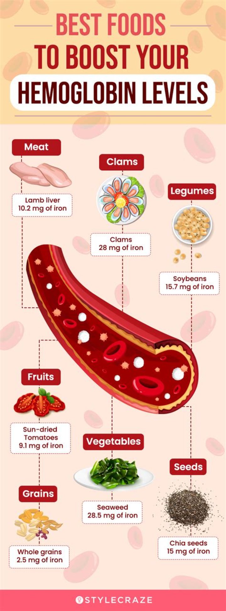
Where are the hepatic veins
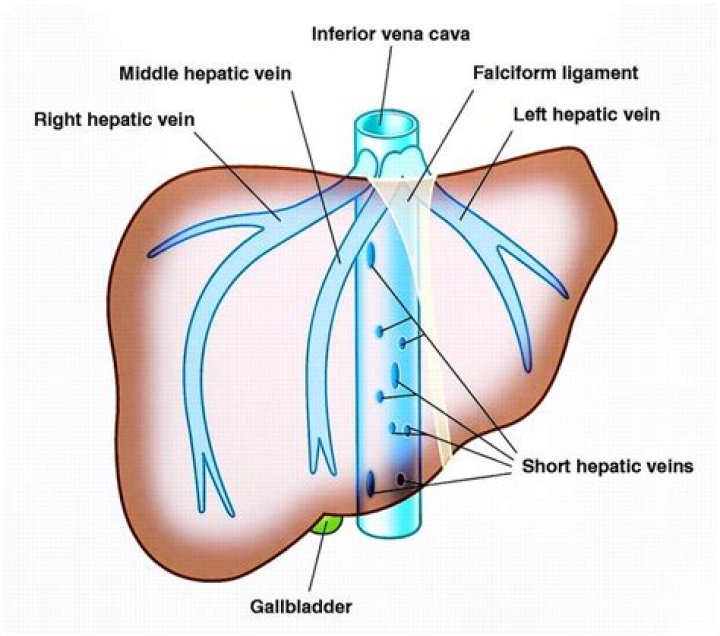
Each hepatic vein can have two or more branches inside the liver. The three main hepatic veins link up at the top of your liver at the inferior vena cava, a large vein that drains the liver to your right heart chamber. On the bottom end of the liver are the organ’s unusual double blood supplies.
What are 3 hepatic veins?
The hepatic veins are three large vessels that drain the venous blood from the liver into the inferior vena cava. The main hepatic veins are the right, intermediate and left hepatic veins. In addition, several smaller and somewhat inconsistent caudate lobe veins contribute to the venous drainage of the liver.
Is central vein and hepatic vein the same?
Central veins of liverFMA71629Anatomical terminology
Where does the hepatic vein enter the liver?
The hepatic portal vein receives blood specifically from the stomach, intestines, pancreas, and spleen, and carries it into the liver through the porta hepatis. The porta hepatis serves as the point of entry for the hepatic portal vein and the proper hepatic artery, and is the point of exit for the bile passages.What organs are drained by the hepatic vein?
Portal veinSystemHepatic portal systemDrains fromGastrointestinal tract, spleen, pancreas
Where do the hepatic veins receive blood from?
The liver receives a blood supply from two sources. The first is the hepatic artery which delivers oxygenated blood from the general circulation. The second is the hepatic portal vein delivering deoxygenated blood from the small intestine containing nutrients.
What is hepatic vein dilated?
Common US findings of CH include a dilated inferior vena cava and dilated hepatic veins (17,18). The right hepatic vein is normally less than 5.6–6.2 mm in diameter at the origin and dilates in response to elevated venous pressure (17). The degree of dilatation correlates with the severity of heart failure (17).
What causes dilated hepatic vein?
Certain medications, diseases, and inherited disorders can cause HVT. Anything that can cause blood clotting in the veins of your liver may lead to HVT. The most common causes of HVT are: inherited blood disorders.How does the hepatic vein divide the liver?
Right hepatic vein divides the right lobe into anterior and posterior segments. Middle hepatic vein divides the liver into right and left lobes (or right and left hemiliver). This plane runs from the inferior vena cava to the gallbladder fossa.
What is the difference between hepatic portal vein and hepatic vein?Hepatic portal vein carries blood and nutrients from the stomach, spleen, intestines and gall bladder to the liver. The hepatic vein carries deoxygenated blood from the liver back to the right atrium of the heart via the inferior vena cava.
Article first time published onWhat is hepatic vasculature?
Abstract. The hepatic circulation is unique in that high volumes of low pressure blood flow are supplied through a dual venous and arterial circulation. This vascular supply is modulated both by the gastrointestinal vascular bed and an intrahepatic microcirculation.
What do hepatic veins do?
The job of the hepatic veins is to move this blood out of your liver. It’s hard work. At any given time, your liver holds about a pint of blood, or about 1/8th of your body’s total blood. The inferior vena cava carries deoxygenated blood from your liver and the lower half of your body to the right side of your heart.
What does hepatic portal vein do?
A blood vessel that carries blood to the liver from the intestines, spleen, pancreas, and gallbladder.
Which vein drains blood from the hepatic flexure of the colon?
The inferior mesenteric vein drains blood from the rectum, sigmoid colon, descending colon and splenic flexure.
How do you access the hepatic vein?
Fluoroscopic approach for obtaining percutaneous hepatic vein access. A, A quadripolar catheter is placed in the coronary sinus through the azygous vein and left superior vena cava. A decapolar catheter is placed in a hepatic vein through the right internal jugular vein.
Where is the left hepatic lobe?
The left lobe is smaller and more flattened than the right. It is situated in the epigastric, and left hypochondriac regions of the abdomen. Its upper surface is slightly convex and is moulded on to the diaphragm; its under surface presents the gastric impression and omental tuberosity.
What is hepatic vein thrombosis?
Hepatic vein thrombosis (Budd-Chiari Syndrome) is a rare disorder resulting from obstruction to the outflow of blood from the liver. The characteristic pathologic findings are intense congestion most pronounced around the terminal hepatic venules, cell necrosis, and a scant inflammatory reaction.
What is the right hepatic lobe?
The right hepatic vein divides the right lobe of the liver into anterior and posterior segments. The middle he- patic vein divides the liver into the right and left lobes and runs in the same plane with the inferior vena cava and the gallbladder fossa.
What happens if the hepatic vein is blocked?
Hepatic vein obstruction prevents blood from flowing out of the liver and back to the heart. This blockage can cause liver damage. Obstruction of this vein can be caused by a tumor or growth pressing on the vessel, or by a clot in the vessel (hepatic vein thrombosis).
What happens if the hepatic portal vein is blocked?
Portal vein thrombosis is blockage or narrowing of the portal vein (the blood vessel that brings blood to the liver from the intestines) by a blood clot. Most people have no symptoms, but in some people, fluid accumulates in the abdomen, the spleen enlarges, and/or severe bleeding occurs in the esophagus.
How do you know if you have a blood clot in your liver?
liver pain. vomiting blood. yellowing of the skin, or jaundice. varices and gastric bleeding.
How many hepatic veins are present?
There are usually three upper hepatic veins draining from the left, middle, and right parts of the liver. These are larger than the group of lower hepatic veins that can number from six to twenty. All of the hepatic veins drain into the inferior vena cava.
Why does the liver have both a hepatic artery and a hepatic portal vein as a blood supply?
The liver is connected to two large blood vessels, the hepatic artery and the portal vein. The hepatic artery carries blood from the aorta to the liver, whereas the portal vein carries blood containing the digested nutrients from the entire gastrointestinal tract, and also from the spleen and pancreas to the liver.
What is the hepatic triad?
por·tal tri·ad. (pōr’tăl trī’ad) Branches of the portal vein, hepatic artery, and the biliary ducts bound together in the perivascular fibrous capsule or portal tract as they ramify within the substance of the liver.
Which vein drains blood from the distal half of the large intestine?
The splenic vein drains the stomach, the superior mesenteric vein drains the upper small intestine, while the inferior mesenteric vein drains the distal portions of the colon. These three tributaries drain into the portal vein, which supplies the liver whose venous effluent is delivered back to the heart [2,4,5].
Where is the Ligamentum Venosum?
Anatomical terminology The ligamentum venosum, also known as Arantius’ ligament, is the fibrous remnant of the ductus venosus of the fetal circulation. Usually, it is attached to the left branch of the portal vein within the porta hepatis. It may be continuous with the round ligament of liver.
Where is the hepatic flexure of the colon?
The right colic flexure or hepatic flexure (as it is next to the liver) is the sharp bend between the ascending colon and the transverse colon. The hepatic flexure lies in the right upper quadrant of the human abdomen. It receives blood supply from the superior mesenteric artery.
What are the 4 main veins draining the stomach?
Portal Venous System Venous drainage from the stomach, duodenum, small bowel, and colon (up to the splenic flexure) is to the superior mesenteric vein. Pancreatic venous drainage is to the splenic and superior mesenteric veins (SMV). The descending and sigmoid colon drain through the inferior mesenteric vein (IMV).
Which part of the large intestine is between the hepatic flexure and the splenic flexure?
ABTransverse ColonThe part of the large intestine between the splenic and hepatic flexures.Descending ColonPart of the large intestine between the splenic flexure and the sigmoid colon.Sigmoid ColonLast part of the large intestine before entry into the rectum.