Where is the articular disc located
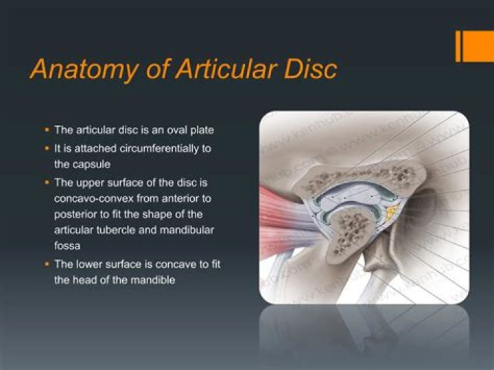
The articular disk is a thin, oval plate, placed between the condyle of the mandible and the mandibular fossa. Its upper surface is concavo-convex from before backward, to accommodate itself to the form of the mandibular fossa and the articular tubercle
Where are articular discs found?
When complete they are called disks; when incomplete they are called menisci. Disks are found in the temporomandibular joint of the lower jaw, the sternoclavicular (breastbone and collarbone) joint, and the ulnocarpal (inner forearm bone and wrist) joint.
What's articular disc?
Medical Definition of articular disk : a cartilage (as the meniscus of the temporomandibular joint) interposed between two articular surfaces and partially or completely separating the joint cavity into two compartments.
Which joint has articular disc?
Articular diskVertical section through the articulations at the wrist, showing the synovial cavities. (Articular disk labeled at center right.)DetailsIdentifiersLatindiscus articularisWhat is the function of the articular disc in the TMJ?
The articular disc in the TMJ has an important functional role. It fills the space between the condyle and the temporal bone, and acts as a stress absorber and distributors during the jaw activity.
Where is the articular disc the thinnest?
Articular disk of the temporomandibular jointTA98A03.1.07.002TA21623FMA57059Anatomical terminology
Does the elbow have an articular disc?
The articular disc is attached to the ulnar styloid by its apex and by its base to the radius. The disc slides over the ulnar end during pronation and supination of the forearm.
Where is TMJ located?
The temporomandibular joints (TMJ) are the 2 joints that connect your lower jaw to your skull. More specifically, they are the joints that slide and rotate in front of each ear, and consist of the mandible (the lower jaw) and the temporal bone (the side and base of the skull).Does shoulder joint have articular disc?
Anatomical terminology The articular disc of the sternoclavicular joint is flat and nearly circular, interposed between the articulating surfaces of the sternum and clavicle.
Does articular cartilage produce synovial fluid?The fluid in articular cartilage effectively serves as a synovial fluid reserve. During movement, the synovial fluid held in the cartilage is squeezed out mechanically to maintain a layer of fluid on the cartilage surface (so-called weeping lubrication).
Article first time published onWhich muscle draws the articular disc of the TMJ forward during opening of the mouth?
The muscles that make direct contact with TMJ are four: masseter, temporal, and two pterygoids. The masseter muscle with its perimysium has direct contact with the articular disc on the front edge.
Is meniscus intra-articular?
Introduction: Menisci and cruciate ligaments are intra-articular structures of knee, and injury to these structures is common.
Do all synovial joints have articular discs?
Many, but not all, synovial joints also contain additional structures: Articular discs or menisci – the fibrocartilage pads between opposing surfaces in a joint. Articular fat pads – adipose tissue pads that protect the articular cartilage, as seen in the infrapatellar fat pad in the knee.
What makes up articular cartilage?
Articular cartilage is hyaline cartilage and is 2 to 4 mm thick. Unlike most tissues, articular cartilage does not have blood vessels, nerves, or lymphatics. It is composed of a dense extracellular matrix (ECM) with a sparse distribution of highly specialized cells called chondrocytes.
What attaches to the articular disc?
Capsule – The capsule is a fibrous membrane that surrounds the joint and attaches to the articular eminence, the articular disc and the neck of the mandibular condyle.
What does articular cartilage look like?
Articular cartilage is the smooth, white tissue that covers the ends of bones where they come together to form joints. Healthy cartilage in our joints makes it easier to move. It allows the bones to glide over each other with very little friction.
Which part of the temporomandibular disc is most avascular?
The central area of the disc is avascular and lacks innervation, thus getting its nutrients from the surrounding synovial fluid. In contrast, the posterior ligament and the surrounding capsules along have both blood vessels and nerves.
Which of the following occur when the articular cartilage is damaged?
Individuals who have severely injured their articular cartilage typically experience the following symptoms: Severe pain and inflammation that increases with activity. Reduced range of motion of the knee joint. Sensation that the knee joints are catching or locking.
What are articular disc made of?
The articular disc of the temporomandibular joint (TMJ) is composed of fibrocartilage, and the extracellular matrix of this disc is composed mainly of collagen, glycosaminoglycan and proteoglycans.
What can be mistaken for TMJ?
- Trigeminal Neuralgia. Just as you have two temporomandibular joints on each side of the face, you also have two trigeminal nerves that control your jaw. …
- Cluster, Migraine, or Tension Headaches. …
- Sinus Issues. …
- Other Causes of TMJ Pain.
How do I realign my TMJ jaw?
Open your mouth as wide as you comfortably can, and hold for 5-10 seconds. Place the tip of your tongue on the roof of your mouth. Glide your lower jaw out as far as it will go and then back in as far as it will go. Hold for 5-10 seconds in each position.
How do you fix uneven jaw clenching?
Take an over-the-counter pain reliever, such as acetaminophen (Tylenol) or ibuprofen (Advil). Avoid strenuous jaw movements. Wear an orthopedic dental appliance to raise your bite and reposition the jaw. Practice TMJ exercises to reduce pain and improve your jaw’s movement.
Does exercise increase synovial fluid?
When a joint moves, the fluid sloshes around, giving the cartilage a healthy dose of oxygen and other vital substances. As an added bonus, regular exercise encourages the body to produce extra synovial fluid. Strong muscles, flexible tendons, and healthy cartilage. These are the things that make everyday life possible.
Where is synovial fluid located in the body?
What is a synovial fluid analysis? Synovial fluid, also known as joint fluid, is a thick liquid located between your joints. The fluid cushions the ends of bones and reduces friction when you move your joints.
What is articular capsule?
In anatomy, a joint capsule or articular capsule is an envelope surrounding a synovial joint. Each joint capsule has two parts: an outer fibrous layer or membrane, and an inner synovial layer or membrane.
What muscles are affected by TMJ?
“When it comes to TMD, we can blame the pain on the masseter muscle, which covers the jaw over your teeth,” says Dr. Bang. “The masseter muscle is used for chewing and jaw clenching. Muscle overuse from teeth grinding and jaw clenching causes the muscles to become tense, inflamed and very painful.”
How long will TMJ pain last?
Acute TMJ symptoms and signs may last anywhere from a few days to a few weeks and then disappear after the injury or cause of discomfort has resolved. For a chronic TMJ condition, the symptoms can be ongoing with episodes of sharp and/or dull pain that occur over an extended period of time (months to years).
Which of the following muscles partially inserts on the articular disc of the temporomandibular joint?
The masticatory muscles are generally described as the muscles that originate from the cranium and insert on the mandible. Some of the masticatory muscles also insert into the articular disc of the temporomandibular joint.
What are the intra-articular structures of knee joint?
The intra-articular ligaments are the anterior cruciate ligament (ACL), posterior cruciate ligament (PCL), and the posterior meniscofemoral ligament. (See the image below.) Knee joint, anterior view. The patellar ligament is the anterior ligament of the knee joint.
What is an extra articular structure of the knee?
Extra articular structures are on the outside or periphery of the articulation between the femur and tibia. Intra-articular structures are in-between femur and tibial area of articulation.
What is intra-articular administration?
An intra-articular injection is a type of shot that’s placed directly into a joint to relieve pain. Corticosteroids (steroids), local anesthetics, hyaluronic acid, and Botox are the most common substances injected into joints for this treatment.