Where is the lateral pterygoid muscle
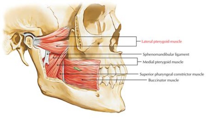
Lateral pterygoid is a two-headed, fan-shaped muscle located in the infratemporal fossa of the skull. It is one of the four masticatory muscles
What is the function of lateral pterygoid muscle?
The Lateral pterygoid muscle is active during mastication and during mandibular movements such as protrusion (forward movement of the mandible), abduction (depression of the mandible), mediotrusion (movement of the mandibular condyle towards the midline), and particularly during speaking, singing, and clenching.
Where do the pterygoid muscles attach?
OriginSuperficial part: Tuberosity of maxilla, Pyramidal process of palatine bone; Deep part: Medial surface of lateral pterygoid plate of sphenoid boneInsertionMedial surface of ramus and angle of mandible
How do you release the lateral pterygoid muscle?
Gently squeeze the muscle between the index finger and the thumb. Start with a gentle pressure, and gradually increase the muscle squeeze as tolerated. Teach the patient to self-squeeze the lateral pterygoid muscle for 1 minute several times per day. Relief of the headache, jaw or facial pain is sometimes immediate.Why is the lateral pterygoid called the peripheral heart?
These communications are important for spread of infections and for collateral circulation. The pterygoid muscles and other muscles of mastication pump the blood from this plexus and are considered a “peripheral heart”. Chewing or yawning increases venous return.
How do you relieve masseter muscle pain?
Heat: Hot packs may be applied to the masseter and jaw to increase circulation, relax the muscle, and decrease pain. Massage: Gentle massage to the masseter may help relax a hypertonic muscle and decrease pain. It may also help improve muscle flexibility.
What muscle lowers the jaw?
masseter: The large muscle which raises the lower jaw, and assists in mastication.
What are the lateral pterygoid muscle attachments?
Attachments of Lateral Pterygoid Muscle: Origin & Insertion Superior head: greater wing of sphenoid bone. … Inferior head: lateral surface of lateral pterygoid plate. Insertion: (distal attachments): Condyle of mandible and temporomandibular joint.What muscles attach to lateral pterygoid plate?
- lateral pterygoid muscle: the lower part of the lateral pterygoid is attached to the lateral aspect of the lateral pterygoid plate.
- medial pterygoid muscle: medial aspect of the lateral pterygoid plate.
- superior pharyngeal constrictor: inferior end of the medial pterygoid plate.
The pterygoid tuberosity is a rough area for the attachment of the medial pterygoid muscle on the internal surface of angle of mandible.
Article first time published onWhich muscle of lower limb is known as peripheral heart?
Also, in upright posture, the soleus is responsible for pumping venous blood back into the heart from the periphery, and is often called the skeletal-muscle pump, peripheral heart or the sural (tricipital) pump.
What muscles elevate and retract the mandible?
The function of the masseter muscle is to elevate the mandible and approximate the teeth—additionally, the intermediate and deep muscle fibers of the masseter function to retract the mandible.
How do soleus muscles act as peripheral hearts?
Together, the calf’s muscles and deep vein system form a complex array of valves and pumps, often referred to as the “peripheral heart,” that functions to push blood upward from the feet against gravity. The calf-muscle pump is analogous to the common hand-pump bulb of a sphygmomanometer filling a blood pressure cuff.
Does the lateral pterygoid muscle elevate the mandible?
The larger deep head originates from the medial surface of the lateral pterygoid plate and the pyramidal process of sphenoid bone. … It receives blood supply from the pterygoid branches of the maxillary artery. The bilateral contraction of this muscle elevates the mandible and closes the mouth.
What muscle moves the jaw from side to side?
The medial pterygoid muscle is innervated by the medial pterygoid branch of the mandibular nerve. Its principal blood supply stems from the pterygoid branches of the maxillary artery. The major functions of this muscle are elevation of the mandible and side-to-side movements when grinding and chewing.
What muscle moves the jaw forward?
The lateral pterygoid muscle pulls the mandible forwards (anterior translatory movement). During this process the mandible moves slightly downwards because the condyle is pressed down on the articular tubercle. The mandible is pulled backwards on closing by the posterior fibres of the temporalis muscle.
How do you examine lateral Pterygoid?
Attempted palpation of what has been thought to be this structure is commonly done by placing the forefinger, or the little finger, over the buccal area of the maxillary third molar region and exerting pressure in a posterior, superior, and medial direction behind the maxillary tuberosity (Figure 2).
How do you palpate the medial and lateral pterygoid?
To palpate from outside the mouth, the head is tilted slightly to access the muscle. Palpation with one finger locates trigger points on the inner surface of the mandible by pressing upward at its angle. Palpation of the mid-belly is performed inside the mouth with the pad of the palpating index finger.
Why does my masseter muscle hurt?
“The masseter muscle is used for chewing and jaw clenching. Muscle overuse from teeth grinding and jaw clenching causes the muscles to become tense, inflamed and very painful.”
Should I massage my masseter muscle?
When massaged properly, it can bring relief from many of the symptoms associated with TMJ syndrome. If over-tightened or clenched, the masseter muscle can cause many complications, such as vertigo, tinnitus, headaches, earaches and toothaches.
Which muscle is attached to Pterygoid fovea?
Background: The pterygoid fovea on the mandibular neck is superomedial to the mandibular notch and serves to attach the lateral pterygoid muscle.
Where does the pterygoid plexus drain into?
Drains fromVenules of the infratemporal fossaDrains toMaxillary vein
Where does the masseter muscle insertion?
The masseter is one of the muscles of mastication. It is a powerful superficial quadrangular muscle originating from the zygomatic arch and inserts along the angle and lateral surface of the mandibular ramus. The masseter is primarily responsible for the elevation of the mandible and some protraction of the mandible.
Which muscle is a mirror image of medial pterygoid?
Medial pterygoidActionselevates mandible, closes jaw, helps lateral pterygoids in moving the jaw from side to sideIdentifiers
Which muscles protract the mandible?
Actions: Acting bilaterally, the lateral pterygoids protract the mandible, pushing the jaw forwards. Unilateral action produces the ‘side to side’ movement of the jaw. Note: Contraction of the lateral pterygoid will produce lateral movement on the contralateral side.
Where is gastrocnemius located?
Gastrocnemius: This muscle is just under your skin at the back of the lower leg. Because the gastrocnemius is close to the skin’s surface, you can often see its outline. It forms the bulk of your calf muscle.
Where are the soleus muscles located?
The soleus muscle, located deep/anterior to the medial and lateral gastrocnemius muscle heads, originates on the posterior aspect of the tibia (middle third of the medial border) and fibula (head and body) and inserts on the calcaneus through the Achilles tendon (see Figure 31.1).
Why do I keep straining my soleus?
While soleus injuries typically result from overuse, a gastrocnemius strain is more likely to be a sudden injury. Commonly called tennis leg, this strain is often the result of a quick movement, such as sprinting or jumping.
What is the pterygoid muscle?
Introduction. The medial pterygoid muscle, a major elevator of the jaw is a square-shaped masticatory muscle, located on the medial aspect of the lower jaw bilaterally. It is also known as internal pterygoid muscle.
What muscle would you use to close your eye?
The orbicularis oculi muscles circle the eyes and are located just under the skin. Parts of this muscle act to open and close the eyelids and are important muscles in facial expression.
Which muscle elevates and retracts the tongue?
The styloglossus originates at the styloid process and blends into the fibers of the inferior longitudinal tongue muscles and the hyoglossus, and elevates and retracts the tongue posteriorly and superiorly.