Which muscle is located laterally to the spine
LUMBAR MUSCLESFUNCTIONQuadratus LumborumLateral flexion of vertebral columnInterspinalesExtends vertebral columnIntertransversarii MedialesLateral flexion of vertebral columnMultifidusExtends & rotates vertebral column
What muscles are next to your spine?
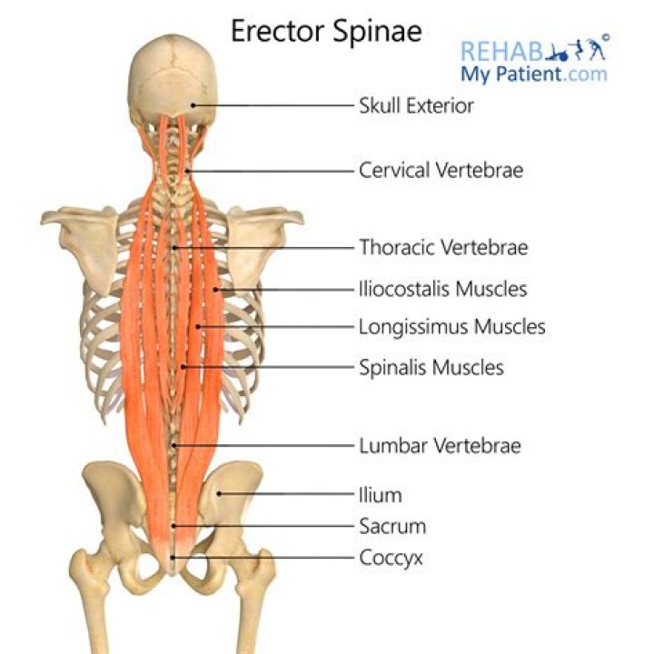
- Iliocostalis muscles. The iliocostalis muscles are those farthest away from your spine. …
- Longissimus muscles. The longissimus muscles help you arch your back and neck. …
- Spinalis muscles. The spinalis muscles are closest to your spine.
What is lateral movement of the spine?
Movement of a body part to the side is called lateral flexion. This type of movement is commonly associated with the neck and spine. For example, when you move your head toward one of your shoulders or bend your body sideways, you’re performing a lateral flexion.
What extends laterally from the spinal cord?
The cord is stabilized within the dura mater by the connecting denticulate ligaments, which extend from the enveloping pia mater laterally between the dorsal and ventral roots. The dural sac ends at the vertebral level of the second sacral vertebra.Where is the paraspinal region?
The paraspinal musculature comprises muscle groups adjacent to the vertebrae and is responsible for the movement and stabilization of the spine. In the lumbar spine, these muscles include the multifidus (MF), erector spinae (ES), interspinales, intertransversarii, psoas major (PM) and quadratus lumborum13,14.
What is the cauda?
Cauda is Latin for tail, and equina is Latin for horse (ie, the “horse’s tail”). The CE provides sensory innervation to the saddle area, motor innervation to the sphincters, and parasympathetic innervation to the bladder and lower bowel (ie, from the left splenic flexure to the rectum).
What is paraspinal muscle spasm?
Lumbar paraspinal muscle spasms are most often clinically diagnosed via a patient history of paraspinal cramps or “knots,” or a finding of splinting, tightness, or decreased range of motion on physical examination.
Which region of the spinal cord contains clusters of cell bodies of somatic motor neurons?
The ventral horns contains the cell bodies of motor neurons that send axons via the ventral roots of the spinal nerves to terminate on striated muscles.Does spinal cord travels adjacent to the spine?
While the vertebral foramen refers to the space in which the spinal cord travels, the intervertebral foramen refers the space between two adjacent vertebrae. The intervertebral foramen allows passage for nerves branching off the spinal cord (spinal nerves) to exit the vertebral foramen and travel to various body areas.
What muscles laterally flex the trunk?The primary muscles involved in lateral flexion of the torso are the internal and external obliques, the quadratus lumborum and the erector spinae. Other muscles — including the rectus abdominis, iliopsoas and semispinalis — assist with the movement.
Article first time published onWhat muscles cause lateral flexion?
All trunk flexors and extensors can produce lateral flexion when acting unilaterally. The major muscles involved are the rectus abdominis, external and internal obliques, erector spinae, semispinalis thoracis, latissimus dorsi, deep posterior spinal muscles, quadratus lumborum, and psoas.
What muscles perform lateral flexion of the vertebral column?
The quadratus lumborum aids in lateral flexion of the vertebral column.
Where are thoracic paraspinal muscles?
The paraspinal muscles are the “action” muscles of the back. When they work, the result is the obvious movement of your spine. They course down your back and spine and help to move your spine into extension, rotation, and side bending.
Which of the following is a paraspinal muscle group?
Erector spinae: Also known as paraspinal muscles, these are a large group of muscles that run from the lower spine to the neck. The muscles from this group specific to the lumbar spine include: Iliocostalis lumborum. Longissimus thoracis.
What does paraspinal mean in medical terms?
Medical Definition of paraspinal : adjacent to the spinal column paraspinal muscles paraspinal tissues.
How do you release paraspinal muscles?
Wrap your arms around your thigh, knee or shin, and gently pull the knee towards your chest. Hold for 20 seconds and slowly extend the leg to starting position. Repeat three times each leg. Use this movement to stretch the paraspinal muscles and strengthen the abdominal muscles.
Which muscle is the large muscle that wraps around the lower back quizlet?
Tendinous intersections. Latissimus dorsi muscle is a lower back muscle. However, the proximal end of the muscle wraps around the torso extending toward the anterior side.
What is cord equina?
The corda equina carries nerves which control the bladder and bowel. The cauda equina also carries nerves which control movement of the legs, and nerves which sense light touch and pain in the legs or around the back passage (perineum).
What is caudal equina?
The cauda equina is the sack of nerve roots (nerves that leave the spinal cord between spaces in the bones of the spine to connect to other parts of the body) at the lower end of the spinal cord. These nerve roots provide the ability to move and feel sensation in the legs and the bladder.
What is Flavum?
One of a series of bands of elastic tissue that runs between the lamina from the axis to the sacrum, the ligamentum flavum connects the laminae and fuses with the facet joint capsules. … As we age, the ligament loses elastin, and this allows the ligament to encroach on the canal.
What vertebrae connects with the spine?
The facet joints link the vertebrae together and give them the flexibility to move against each other. Each vertebra has a hole in the center, so when they stack on top of each other they form a hollow tube that holds and protects the entire spinal cord and its nerve roots.
Where is T2 located in the spine?
The T2 vertebra is a member of the thoracic vertebrae column, located between the cervical vertebrae and the lumbar spinal vertebrae. As the second descending thoracic vertebra, the T2 vertebra is located below T1 and above T3.
Where does the thoracic spine start and end?
The thoracic region of the spine begins at the base of your neck and ends around the bottom of your rib cage, just above your lower back. The thoracic region of the spine, the upper back, is located below the cervical region (neck) and above the lumbar region (lower back).
Which region of the spinal cord contains clusters of cell bodies of somatic motor neurons quizlet?
1. Each region of the gray matter has very specific functions. a. Anterior gray horn: Contains the cell bodies of somatic motor neurons.
Where is lateral horn of spinal cord?
The lateral horn of the spinal cord is the small lateral projection of grey matter located between the dorsal horn and ventral horn and contain the neuronal cell bodies of the sympathetic nervous system.
Where are interneurons located in the spinal cord?
Most interneurons are found in the grey column, a region of grey matter in the spinal cord.
Which muscle is located posterior to the spine?
These deep muscles are enclosed by fascia. The deep back muscles are posterior to the erector spinae. They are short muscles associated with the spinous and transverse processes of the vertebrae. The three deep muscles of the back include the semispinalis, multifidus, and rotatores.
What is lateral flexion of the lumbar spine?
Lumbar Spine. Lateral flexion is side bending in the frontal/ coronal plane. The quadratus lumborum, oblique abdominals, and erector spinae are considered the primary lateral flexors. Note that the internal and external obliques on the same side work to produce the same motion.
What are hip flexors muscles?
Your hip flexors are a group of muscles near the top of your thighs that are key players in moving your lower body. They let you to walk, kick, bend, and swivel your hips. But if your muscles are too tight or if you make a sudden movement, your hip flexors can stretch or tear.
What muscle flexes and laterally flexes the trunk?
Quadratus lumborum is involved in extending the trunk and flexing it laterally to the ipsilateral side.
What muscles do side bends?
Side bends primarily target your obliques, which are the muscles that wrap around your waist and torso.