What is parabasal cells on Pap smear
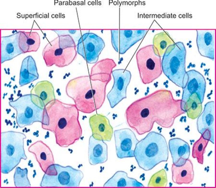
Parabasal cells are the smallest epithelial cells seen on a typical vaginal smear
What causes parabasal cells?
At the cytologic level, fewer superficial epithelial cells are observed, but an increased percentage of intermediate and parabasal cells are found as a result of estrogen deficiency.
What is atrophic pattern in pap smear?
Sometimes after menopause or when breast feeding there are ‘atrophic changes’ in the cervix, caused by decreased hormone levels. If your Pap smear result is ‘atrophic’ you may be given some local oestrogen treatment (for example, oestrogen cream) and asked to have the Pap smear repeated in three months.
What are intermediate cells?
Intermediate cells – two to three times larger than parabasal cells, but their nuclei are similar in size to parabasal cell nuclei. Small intermediate cells are round or almost round with a large prominent nucleus. … The nuclei are absent or pyknotic, meaning small and dark.What does it mean when endocervical cells are present?
Endocervical cells present. This phrase means that cells from the inside of your cervical canal were sampled at the time of the pap test, which is something your doctor tries to do.
What are nucleated epithelial cells?
Nucleated epithelial cells are round or oval-shaped cells, larger than polymorphonuclear cells, that have a clear cytoplasm and a centrally located nucleus that stains strongly with methylene blue. Cornified epithelial cells are the largest cells in the smear. They are flattened and angular in outline.
What can cause inflammation on a Pap smear?
Inflammation on a Pap smear can be found in a patient of any age and may be attributable to a benign infection — such as Candida vaginitis — that need be treated only if the patient is symptomatic. Sexually transmitted infections can also cause an inflammatory reaction on the cervix and should be treated accordingly.
What kind of cells predominates in vaginal smear during Metestrus stage?
The cell types present in vaginal smears during this stage are fragmented, cornified epithelial cells and smaller darker stained leukocytes (Figure 1F, Figure 2, Metestrus, right panel).When are intermediate cells seen?
Intermediate Squamous Cells The intermediate cell’s cytoplasm is thin, transparent, and typically stains basophilic. The centrally placed nucleus is 35 µm. The nucleus is vesicular with fine evenly dispersed granular chromatin. Intermediate squames are seen in abundance when progesterone is at high levels.
What is the main function of intermediate filaments?Intermediate filaments are therefore found in particularly durable structures such as hair, scales and fingernails. The primary function of intermediate filaments is to create cell cohesion and prevent the acute fracture of epithelial cell sheets under tension.
Article first time published onWhat does vaginal atrophy feel like?
Vaginal atrophy (also called atrophic vaginitis) is a condition where the lining of the vagina gets drier and thinner. This results in itching, burning and pain during sex, among other symptoms. The condition also includes urinary tract problems such as urinary tract infections (UTIs) and frequent urination.
What does atrophic mean?
atrophy, decrease in size of a body part, cell, organ, or other tissue. The term implies that the atrophied part was of a size normal for the individual, considering age and circumstance, prior to the diminution.
How do you treat atrophy naturally?
- Herbs and supplements. Researchers in a 2014 study examined sea buckthorn oil as a possible alternative to traditional estrogen therapy. …
- Diet. Reaching and maintaining a healthy weight and body mass index may also help with GSM. …
- Exercise. …
- Personal care products.
Are endocervical cells cancerous?
Cell types This is called the endocervix. The skin-like cells of the ectocervix can become cancerous, leading to a squamous cell cervical cancer. This is the most common type of cervical cancer. The glandular cells of the endocervix can also become cancerous, leading to an adenocarcinoma of the cervix.
What is the difference between endocervical and cervical?
The cervix is made of two parts and is covered with two different types of cells. The endocervix is the opening of the cervix that leads into the uterus. It is covered with glandular cells. The exocervix (or ectocervix) is the outer part of the cervix that can be seen by the doctor during a speculum exam.
What is HPV high risk positive?
A positive test result means that you have a type of high-risk HPV that’s linked to cervical cancer. It doesn’t mean that you have cervical cancer now, but it’s a warning sign that cervical cancer could develop in the future.
How common is inflammation on PAP?
Ten percent had inflammation on their Pap smear; these women were more likely than those without inflammation to test positive for chlamydia and trichomonas. However, 71 percent of women with inflammation had no evidence of any organisms.
What are the 5 classic signs of inflammation?
Five cardinal signs characterize this response: pain, heat, redness, swelling, and loss of function.
How do you treat an inflammatory smear?
If you have cervical inflammation due to cervical cancer or precancer, you doctor may perform cryosurgery, freezing abnormal cells in the cervix, which destroys them. Silver nitrate can also destroy abnormal cells. Your doctor can treat your cervicitis after they know its cause.
What is epithelial cells in the vagina?
The vaginal epithelium is the inner lining of the vagina consisting of multiple layers of (squamous) cells. The basal membrane provides the support for the first layer of the epithelium-the basal layer. The intermediate layers lie upon the basal layer, and the superficial layer is the outermost layer of the epithelium.
What is epithelial cell abnormality?
Epithelial cell abnormalities This means that the cells lining the cervix or vagina show changes that might be cancer or a pre-cancer. This category is divided into several groups for squamous cells and glandular cells.
What are epithelial cells squamous?
Squamous epithelial cells are large, polygonal cells with small round nuclei. They tend to fold on themselves and sometimes are confused with casts. Their large size allows them to be easily distinguished from casts. (2) Common in voided or catheterized samples due to urethral or vaginal contamination.
How do I read my pap smear results?
- Normal. A normal (or “negative”) result means that no cell changes were found on your cervix. …
- Unclear (ASC-US) It is common for test results to come back unclear. …
- Abnormal. An abnormal result means that cell changes were found on your cervix. …
- Negative. …
- Positive.
Are squamous cells normal in Pap smear?
A normal Pap smear shows healthy squamous cells (flat cells that look like fish scales) from the surface of the cervix. There are no signs of infection and no abnormal cells.
What does atypical squamous cells of undetermined significance?
Atypical squamous cells of undetermined significance is the most common abnormal finding in a Pap test. It may be a sign of infection with certain types of human papillomavirus (HPV) or other types of infection, such as a yeast infection.
What is k9 cytology?
Cytology provides a quick and minimally invasive way to evaluate cutaneous tumors in dogs and cats. It may provide a definitive diagnosis and can, thus, help the clinician to determine whether further staging tests, such as lymph node aspiration or chest radiography, are indicated.
What do Cornified cells look like?
Six types of cells typify vaginal cytology: parabasal cells (A), which look like small, O-shaped oat cereal pieces; small intermediate and large intermediate cells (B), which look like fried eggs; superfi- cial (“cornified”) (C) cells, which look like corn flakes; neutrophils; and red blood cells.
What is the difference between menstrual and estrous cycle?
The key difference between estrous and menstrual cycle is that estrous cycle is the reproductive cycle of females of non-primate mammals in which the endometrium is reabsorbed by the walls of the uterus while menstrual cycle is the reproductive cycle of females of primate mammals in which endometrium is shed via …
What is the difference between microfilaments and intermediate filaments?
Microfilaments are often associated with myosin. They provide rigidity and shape to the cell and facilitate cellular movements. Intermediate filaments bear tension and anchor the nucleus and other organelles in place.
Which cellular function may be disrupted because of a malformation of the intermediate filaments?
Intermediate filaments are structural proteins. Microfilaments are used for movement and contain the actin filaments used in muscle contraction. Which cellular function may be disrupted because of a malformation of the intermediate filaments? Mutations can change the overall structure of proteins.
Why are intermediate filaments called intermediate filaments?
Initially designated ‘intermediate’ because their average diameter (10 nm) is between those of narrower microfilaments (actin) and wider myosin filaments found in muscle cells, the diameter of intermediate filaments is now commonly compared to actin microfilaments (7 nm) and microtubules (25 nm). …