What is seen in hypertensive retinopathy
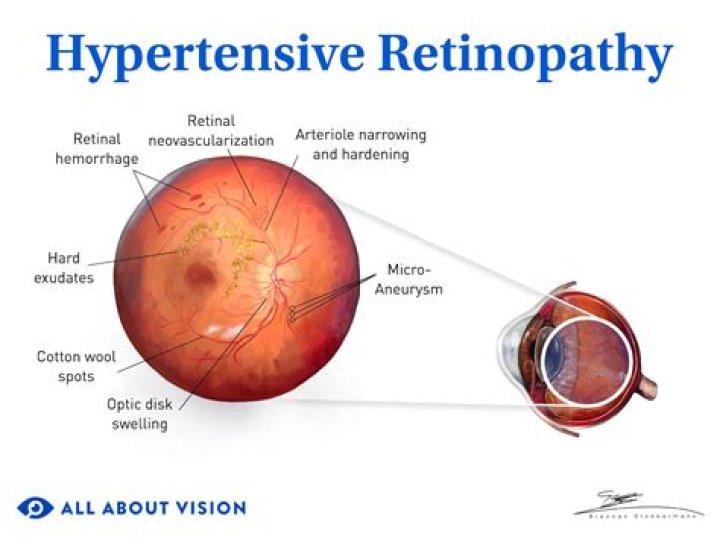
Hypertensive retinopathy is retinal vascular damage caused by hypertension. Signs usually develop late in the disease. Funduscopic examination shows arteriolar constriction, arteriovenous nicking, vascular wall changes, flame-shaped hemorrhages, cotton-wool spots
Is Papilledema seen in hypertensive retinopathy?
On fundoscopic exam, flame and dot blot hemorrhages, hard exudates, cotton wool spots, retinal edema, and papilledema (present in severe hypertensive retinopathy) can be seen.
How can you tell the difference between hypertensive and diabetic retinopathy?
Both cause damage to the retina, but they have different causes. Diabetic retinopathy is caused by high blood sugar. Hypertensive retinopathy is caused by high blood pressure. Both conditions are diagnosed by an eye doctor.
What do cotton wool spots indicate?
Cotton-wool spots (CWSs) are common retinal manifestations of many diseases including diabetes mellitus, systemic hypertension, and acquired immunodeficiency syndrome. Clinically they appear as whitish, fluffy patches on the retina and eventually fade with time.Does neovascularization occur in hypertensive retinopathy?
Hypertensive retinopathy can be rarely complicated with retinal neovascularization. Treatment with PRP can be undertaken.
Are flame hemorrhages seen in hypertensive retinopathy?
Signs of hypertensive retinopathy include superficial, flame-shaped hemorrhages, arteriovenous crossing changes (nicking), retinal arteriole narrowing/straightening, copper- or silver-wire arteriole changes (arteriolosclerosis), cotton-wool spots, microaneurysms, hard exudates (may be in a circinate or macular star …
Are exudates seen in hypertensive retinopathy?
The signs of hypertensive retinopathy include constricted and tortuous arterioles, retinal hemorrhage (Figure 1-3), hard exudates (Figure 2), cotton wool spots (Figure 1 & 3), retinal edema, and papilledema (Figure 3).
What are retinal exudates?
Retinal edema and hard exudates are caused by the breakdown of the blood-retina barrier, allowing leakage of serum proteins, lipids, and protein from the vessels.What is drusen?
Drusen are small, yellowish deposits of cellular debris that accumulate under the retina — the light-sensitive layer of cells at the back of the eye that’s essential to vision. Drusen occur in most people over age 60 and are more common in women than men.
What's the difference between exudates and drusen?Exudates are caused by leaking fatty deposits from blood vessels and appear in compact groups, whereas drusen are believed to be a result of a reduced capacity of the retina to cleanse waste products from the photoreceptors and can appear over the whole retina.
Article first time published onHow does high blood pressure cause diabetic retinopathy?
Increased blood pressure has been hypothesised, through the effects of increased blood flow, to damage the retinal capillary endothelial cells in eyes of people with diabetes.
What is copper wiring in the eye?
Initially, the increased thickness of the vessel walls causes the reflex to be more diffuse and less bright. Progression of sclerosis and hyalinization causes the reflex to be more diffuse and the retinal arterioles to become red-brown. This is known as copper wiring.
What are the stages of diabetic retinopathy?
When these blood vessels thicken, they can develop leaks, which can then lead to vision loss. The four stages of diabetic retinopathy are classified as mild, moderate, and severe nonproliferative and proliferative.
Is ocular hypertension the same as hypertensive retinopathy?
OCULAR COMPLICATIONS OF HYPERTENSION High blood pressure can damage blood vessels in the retina. Hypertensive retinopathy is damage to the retina due to a systemic blood pressure higher than the eye can tolerate.
What is silver wiring in the eye?
Silver wiring or copper wiring is where the walls of the arterioles become thickened and sclerosed causing increased reflection of the light. Arteriovenous nipping is where the arterioles cause compression of the veins where they cross. This is again due to sclerosis and hardening of the arterioles.
What is silver wiring?
Retinal arterioles appear orange or yellow instead of red (“copper wiring” ) Retinal arterioles look white if they have become occluded (“silver wiring” ) Retinal arterioles indent retinal veins as they cross each other (“arteriovenous nicking” )
What are flame hemorrhages?
Flame hemorrhages are a subset of retinal hemorrhages occurring within the retinal nerve fiber layer. 1. They are related to pathologies of the superficial retinal capillary plexus,1 including hypertensive retinopathy and retinal venous occlusion.
What are dot and blot hemorrhages?
Dot and blot hemorrhages occur as microaneurysms rupture in the deeper layers of the retina, such as the inner nuclear and outer plexiform layers. These appear similar to microaneurysms if they are small; fluorescein angiography may be needed to distinguish between the two.
What is microaneurysms of the eye?
Definition. Microaneurysms are tiny outpouchings of blood that protrude from an artery or vein. When they occur in the eye, they are known as retinal microaneurysms. If these protrusions open, they leak blood into the tissues of the retina.
What causes PED?
The pathogenesis of PED formation in AMD is not completely understood, but it is believed to be due to the growth of choroidal neovascularization through Bruch’s membrane into the sub-RPE space with secondary extravasation of fluid or blood.
What is the difference between hard and soft drusen?
Hard drusen are small and round, have well-defined borders and are often spread out. They are common as people age. Soft drusen are larger, have indistinct borders and tend to cluster together. Although both types of drusen should be monitored, hard drusen don’t usually cause vision problems.
What is dry macular?
Dry macular degeneration is a common eye disorder among people over 50. It causes blurred or reduced central vision, due to thinning of the macula (MAK-u-luh). The macula is the part of the retina responsible for clear vision in your direct line of sight.
What is Circinate pattern?
Circular; ring-shaped.
What is exudate and transudate?
“Transudate” is fluid buildup caused by systemic conditions that alter the pressure in blood vessels, causing fluid to leave the vascular system. “Exudate” is fluid buildup caused by tissue leakage due to inflammation or local cellular damage.
What causes peripheral retinal drusen?
Ocular conditions that cause drusen include: Age-related macular degeneration (AMD) and its variants, familial dominant drusen, Best vitteliform macular dystrophy and optic nerve drusen.
What is the outermost layer of the retina?
The center of the macula is called the fovea. The inner surface of the retina is adjacent to the vitreous of the eye. The outermost layer of the retina, the retinal pigment epithelium, is tightly attached to the choroid.
Is drusen macular degeneration?
Drusen are the defining feature of macular degeneration. These small yellow or white spots on the retina can be detected by an ophthalmologist during a dilated eye exam or with retinal photography. People with more than a few small drusen are said to have early age-related macular degeneration (AMD).
What is inflammatory exudate?
Exudate is fluid that leaks out of blood vessels into nearby tissues. The fluid is made of cells, proteins, and solid materials. Exudate may ooze from cuts or from areas of infection or inflammation. It is also called pus.
How is hypertensive retinopathy diagnosed?
Diagnosing hypertensive retinopathy typically involves an examination by an ophthalmologist based on the symptoms present. In some cases, an ophthalmoscope may be used to investigate the retina in the back of the eye. This instrument shines light into the eye, allowing doctors to see any signs of damage.
Does high blood pressure affect diabetic retinopathy?
Systolic, diastolic, and pulse blood pressures did not correlate with blood glucose level in any of our patients. These data revealed that systolic hypertension is a risk for diabetic retinopathy, especially for blot hemorrhage.
Does high blood pressure cause high eye pressure?
High Blood Pressure and Glaucoma Doctors know that increased blood pressure results in increased eye pressure, possibly because high blood pressure increases the amount of fluid the eye produces and/or affects the eye’s drainage system.