What is the lamina Cribrosa
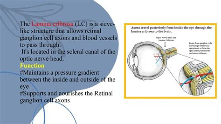
The lamina cribrosa (LC) is a reticulated, sieve-like structure that fills the posterior scleral foramen, which unmyelinated retinal ganglion cell (RGC) axons pass through before converging as the optic nerve (ON).
Where is the lamina cribrosa?
The lamina cribrosa forms the bottom of the optic cup on the inner surface of the optic nerve head. On the outer surface of the optic nerve head, the posterior part of the lamina cribrosa faces the anterior region of the optic nerve.
What passes through the lamina cribrosa?
Lamina cribrosa sclerae, a mesh-like structures which allows nerve fibres of the optic nerve to pass through the sclera.
What is the lamina cribrosa of sclera?
The nerve fibers forming the optic nerve exit the eye posteriorly through a hole in the sclera that is occupied by a mesh-like structure called the lamina cribrosa. It is formed by a multilayered network of collagen fibers that insert into the scleral canal wall.What is human lamina?
Abstract. The lamina cribrosa is a sieve-like perforation in the posterior part of the sclera, that allows passage of the retinal ganglion cell axons and central retinal vessels and preserves a pressure gradient between the intraocular and extraocular space.
Does the lamina Cribrosa have blood vessels?
The anterior part of lamina cribrosa is vascularized by choroidal vessels and in a minor part by the vascular ring of Zinn-Haller.
What is area Cribrosa?
The apical portion called the papilla, of each renal pyramid projects into a minor calyx. The tip of the papilla is perforated with the openings of 10-25 papillary ducts, the final collecting ducts of the uriniferous tubules, and is therefore called the area cribrosa.
What is located in the fovea?
Fovea: In the eye, a tiny pit located in the macula of the retina that provides the clearest vision of all. Only in the fovea are the layers of the retina spread aside to let light fall directly on the cones, the cells that give the sharpest image. Also called the central fovea or fovea centralis.What is unspecified Papilledema?
Papilledema is a serious medical condition where the optic nerve at the back of the eye becomes swollen. Symptoms can include visual disturbances, headaches, and nausea. Papilledema occurs when there is a buildup of pressure in or around the brain, which causes the optic nerve to swell.
Where is the fundus of the eye?Fundus is the bottom or base of anything. In medicine, it is a general term for the inner lining of a hollow organ. The ocular fundus is the inner lining of the eye made up of the Sensory Retina, the Retinal Pigment Epithelium, Bruch’s Membrane, and the Choroid.
Article first time published onWhat artery passes through optic canal?
The optic canal is a very important structure due to the structures that pass through this canal, mainly the optic nerve and the ophthalmic artery.
What is ora serrata in human eye?
The ora serrata is the peripheral termination of the retina and lies approximately 5 mm anterior to the equator of the eye. … The ora serrata is approximately 2 mm wide and is the site of transition from the complex, multilayered neural retina to the single, nonpigmented layer of ciliary epithelium.
Is the white of the eye?
ScleraTA26750FMA58269Anatomical terminology
What is a medullary ray kidney?
The medullary rays are well-defined anatomic structures consisting of bundles of renal tubules which form in the renal cortex and continue through the renal medulla as the medullary striations. … Under these conditions, fine striations are visualized which correspond in position and orientation to these structures.
What is medullary rays in plants?
Medullary rays are strips of parenchyma present between vascular bundles of dicot stem. They separate xylem and phloem bundles. They serve as a link between pith and cortex. They are also known as pith rays and vascular rays.
What is lobule of kidney?
A cortical lobule (or renal lobule) is a part of a renal lobe. It consists of the nephrons grouped around a single medullary ray, and draining into a single collecting duct. Its near identical parallel is the rectal lobe, which is present in the majority of mammals.
What is ocular Hypotony?
Hypotony is usually defined as an intraocular pressure (IOP) of 5 mm Hg or less. Low IOP can adversely impact the eye in many ways, including corneal decompensation, accelerated cataract formation, maculopathy, and discomfort. Clinically significant changes occur more frequently as the IOP approaches 0 mm Hg.
What is circle of Zinn Haller?
The circle of Haller and Zinn comprises complete or incomplete anastomoses around the optic nerve between the medial and lateral short posterior ciliary arteries (SPCAs), which form a dense capillary plexus around the optic nerve.
What is a Cilioretinal artery?
Cilioretinal arteries arise from the short posterior ciliary arteries, as does the choroidal circulation. They make a characteristic bend as they leave the disc margin and are recognisable on fundoscopy.
What is the most common cause of papilledema?
The most common causes of papilledema without IIH were intracranial tumor, intracranial hemorrhage, and cerebral venous sinus thrombosis (Table 1).
What are the symptoms of papilledema?
Symptoms of Papilledema Fleeting vision changes—blurred vision, double vision, flickering, or complete loss of vision—typically lasting seconds are characteristic of papilledema. Other symptoms may be caused by the elevated pressure in the brain. Headache, nausea, vomiting, or a combination may occur.
What medications can cause papilledema?
- Corticosteroids.
- Isotretinoin.
- Lithium.
- Tetracycline.
What is macula and fovea?
The macula is the pigmented part of the retina located in the very center of the retina. In the center of the macula is the fovea, perhaps the most important part of the eye. The fovea is the area of best visual acuity. It contains a large amount of cones—nerve cells that are photoreceptors with high acuity.
What is fovea psychology?
a small depression in the central portion of the retina in which retinal cone cells are most concentrated and an image is focused most clearly. Also called fovea.
What is the fovea and its function?
The fovea is responsible for sharp central vision (also called foveal vision), which is necessary in humans for activities for which visual detail is of primary importance, such as reading and driving.
What is a fundus eye?
The fundus is the inside, back surface of the eye. It is made up of the retina, macula, optic disc, fovea and blood vessels. With fundus photography, a special fundus camera points through the pupil to the back of the eye and takes pictures. These pictures help your eye doctor to find, watch and treat disease.
What is the Fundus examination?
Ophthalmoscopy is a test that allows your ophthalmologist, or eye doctor, to look at the back of your eye. This part of your eye is called the fundus, and consists of: retina. optic disc. blood vessels.
What is a normal fundus?
Normal Fundus. The disk has sharp margins and is normal in color, with a small central cup. Arterioles and venules have normal color, sheen, and course. Background is in normal color. The macula is enclosed by arching temporal vessels. The fovea is located by a central pit.
Where is sphenoid bone?
An unpaired bone located in the cranium (or skull), the sphenoid bone, also known as the “wasp bone,” is located in the middle and toward the front of the skull, just in front of the occipital bone.
Does the sphenoid bone contain a sinus?
Also, several fissures and foramina exist in the sphenoid, which transports several blood vessels and nerves of the skull to the head and neck. The body of the sphenoid has a cavity with a sinus that communicates with the nasal cavity.
What foramen does the oculomotor nerve pass through?
Cranial NerveForamenComponentsII-Opticoptic canal of sphenoidspecial sensoryIII-Oculomotorsuperior orbital fissuresomatomotorvisceromotorIV-Trochlearsuperior orbital fissuresomatomotor