What type of cell contains rhodopsin
Rhodopsin is the visual pigment of the rod photoreceptor
What type of cells is rhodopsin found in?
Rhodopsin is found in specialized light receptor cells called rods. As part of the light-sensitive tissue at the back of the eye (the retina), rods provide vision in low light.
Do cones contain rhodopsin?
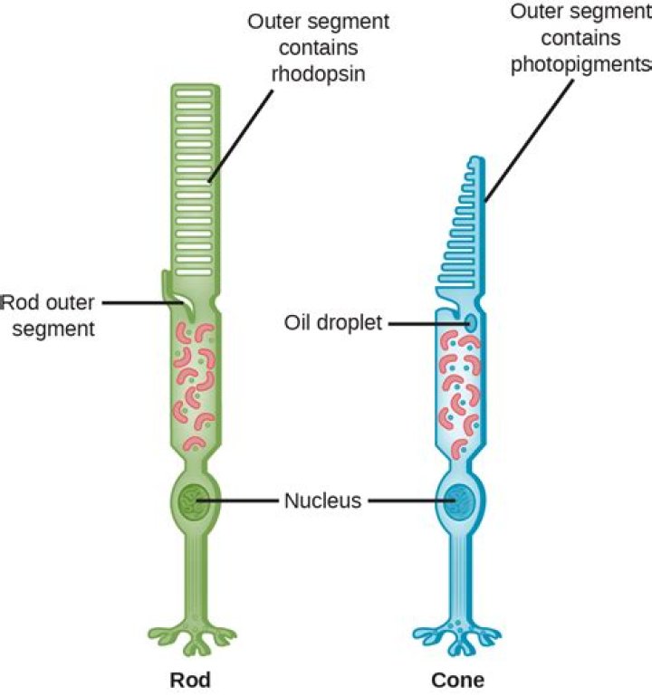
These pigment proteins belong to a family known as the opsins. The pigment protein in rods is called rhodopsin, while the pigment protein in cones is called iodopsin. A single rod can contain up to 100 million molecules of rhodopsin in its outer segment discs.
What part of the eye contains rhodopsin?
Rhodopsin is a biological pigment found in the rods of the retina and is a G-protein-coupled receptor (GPCR). It belongs to a group of photoswitchable opsins. Rhodopsin is extremely sensitive to light, and thus enables vision in low-light conditions.Is rhodopsin in cones and rods?
Introduction. In the retinas of most vertebrates, there are two types of photoreceptor cells, rods and cones (Fig. … Rods contain a single rod visual pigment (rhodopsin), whereas cones use several types of cone visual pigments with different absorption maxima.
Is rhodopsin a receptor?
Rhodopsin is the light receptor in rod photoreceptor cells of the retina that initiates scotopic vision. In the dark, rhodopsin is bound to the chromophore 11-cis retinal, which locks the receptor in an inactive state.
What molecules make up rhodopsin?
Structurally, rhodopsin is classified as a chromoprotein (chromo is a Greek-derived root meaning “colour”). It is made up of opsin (a colourless protein) and 11-cis-retinal (11-cis-retinaldehyde), a pigmented molecule derived from vitamin A.
Is rhodopsin an enzyme?
Rhodopsin phosphodiesterase (Rh-PDE) is an enzyme rhodopsin belonging to a recently discovered class of microbial rhodopsins with light-dependent enzymatic activity.Where is rhodopsin made?
Rhodopsin is synthesized in the endoplasmic reticulum and passes to the Golgi membranes where it becomes glycosylated. Rhodopsin-containing vesicles move from the Golgi to the outer segment where they fuse with the outer segment plasma membrane.
What chromosome is the RHO gene on?The death of cone cells leads to tunnel vision and ultimately blindness in many affected individuals. It is unclear how mutations in the RHO gene affect the function and survival of cone cells. The RHO gene is found on chromosome 3 ( me/3/).
Article first time published onWhat is a ganglion cell?
Ganglion cells are the final output neurons of the vertebrate retina. Ganglion cells collect information about the visual world from bipolar cells and amacrine cells (retinal interneurons). This information is in the form of chemical messages sensed by receptors on the ganglion cell membrane.
What are bipolar cells?
Bipolar cells are the only neurons that connect the outer retina to the inner retina. They implement an ‘extra’ layer of processing that is not typically found in other sensory organs.
What is located in the fovea?
Fovea: In the eye, a tiny pit located in the macula of the retina that provides the clearest vision of all. Only in the fovea are the layers of the retina spread aside to let light fall directly on the cones, the cells that give the sharpest image. Also called the central fovea or fovea centralis.
What is rhodopsin function?
Rhodopsin is a G-protein coupled receptor, and is the most abundant protein in the rod cells found in the retina (Figure 1). It functions as the primary photoreceptor molecule of vision, and contains two parts: an opsin molecule linked to a chromophore, 11-cis-retinal (Athanasiou et al., 2018).
Is rhodopsin AG protein coupled receptor?
Crystal structure of rhodopsin: A G protein-coupled receptor.
Are rods found in the fovea?
In the fovea, there are NO rods… only cones. The cones are also packed closer together here in the fovea than in the rest of the retina. Also, blood vessels and nerve fibers go around the fovea so light has a direct path to the photoreceptors.
How many amino acids make up rhodopsin?
Bovine rhodopsin exists physiologically as ahomodimer consisting of 349 amino acids each and lies near the middle of the spectrum of polypeptide chain lengths among the GPCR family (1). The molecular weight of each distinct peptide chain is 39,119 Da, and the isoelectric point (pI) is 5.88 (11).
How does retina bind to rhodopsin?
One function is to bind retinal. Rhodopsin is a protein that is essential for vision, especially in dim light. The photoreceptors in the retina that contain rhodopsin are rods. Rhodopsin is attached to 11-cis retinal which becomes excited by a photon of light and isomerizes to become all-trans conformation.
How many amino acids are in rhodopsin?
The amino acid sequence of human rhodopsin, deduced from the nucleotide sequence of its gene, is 348 residues long and is 93.4% homolo- gous to that of bovine rhodopsin.
Is rhodopsin a transport protein?
A. Rhodopsin transport carriers (RTCs) that replenish the ROS sensory membrane are detected by EM immunocytochemistry in the rod inner segment (RIS), at the base of the cilium (C) (also called the connecting cilium) of Rana Berlandieri photoreceptors.
Does rhodopsin have quaternary structure?
Rhodopsin is a prototypical G-protein coupled receptor that initiates photo-transduction in the retina of the eye. … Our results suggest that the quaternary structure of wild-type rhodopsin is vastly different compared to that of the misfolded mutant rhodopsin.
What is true rhodopsin?
rhodopsin (visual purple) (roh-dop-sin) n. a pigment in the retina of the eye consisting of retinal and a protein. The presence of rhodopsin is essential for vision in dim light.
Does rhodopsin have beta sheets?
The molecular weight of the homodimer is 78,238 Da. The structure of rhodopsin is intrinsically linked to its function. … Helices constitute the majority (60%, 211 residues) of rhodopsin’s secondary structure, while beta-sheets account for only 2% (10 residues).
What is bovine rhodopsin?
Rhodopsin is the visual pigment that mediates dim-light vision in vertebrates and is a model system for the study of retinal disease. … Keywords: opsin; retinal disease; retinitis pigmentosa; vision; visual pigment.
Where is rhodopsin kinase found?
Rhodopsin kinase is found primarily in mammalian retinal rod cells, where it phosphorylates light-activated rhodopsin, a member of the family of G protein-coupled receptors that recognizes light.
How is rhodopsin activated?
This is consistent with the observation that rhodopsin is activated by the photon-triggered isomerization of retinal in the ligand binding pocket, which requires the ligand not only being bound but also being tightly hold in the pocket, while most other GPCRs are activated by simply binding to the ligands.
What animals have rhodopsin?
In this study we present the most detailed comparative phylogenetic study of mammal rhodopsins to date. We include several groups that are highly specialized for living in low light conditions, including bats, subterranean mole-rats, pinnipeds and cetaceans.
What chromosome is rhodopsin on?
The human rhodopsin gene has been assigned to human chromosome 3 through the use of a mouse DNA probe and human/mouse somatic cell hybrids.
What is the gene locus for rhodopsin?
Japanese RHO locus is comprised of eight major haplotypes. Retinitis pigmentosa-associated haplotype was not identified. Frequency and pattern of rhodopsin point mutations in Chinese patients with autosomal dominant retinitis pigmentosa. The eye photoreceptor protein rhodopsin.
What is Iodopsin and rhodopsin?
Rhodopsin is light absorbing pigment (rhodopsin) present inside rod cells of humans for night vision. Iodopsin is violet color pigment in cones of chicken eyes for color vision. Iodopsin is close analogue of visual purple rhodopsin that is used in night vision.
What is amacrine cells?
Amacrine cells are interneurons in the retina. … Amacrine cells are inhibitory neurons, and they project their dendritic arbors onto the inner plexiform layer (IPL), they interact with retinal ganglion cells and/or bipolar cells.